. 2023 Feb 24;24(1):67.
doi: 10.1186/s12859-023-05175-6.

Immunoinformatics-aided design of a new multi-epitope vaccine adjuvanted with domain 4 of pneumolysin against Streptococcus pneumoniae strains

Affiliations

Immunoinformatics-aided design of a new multi-epitope vaccine adjuvanted with domain 4 of pneumolysin against Streptococcus pneumoniae strains

Mona Shafaghi et al. BMC Bioinformatics. .

Abstract

Background: Streptococcus pneumoniae (Pneumococcus) has remained a leading cause of fatal infections such as pneumonia, meningitis, and sepsis. Moreover, this pathogen plays a major role in bacterial co-infection in patients with life-threatening respiratory virus diseases such as influenza and COVID-19. High morbidity and mortality in over one million cases, especially in very young children and the elderly, are the main motivations for pneumococcal vaccine development. Due to the limitations of the currently marketed polysaccharide-based vaccines, non-serotype-specific protein-based vaccines have received wide research interest in recent years. One step further is to identify high antigenic regions within multiple highly-conserved proteins in order to develop peptide vaccines that can affect various stages of pneumococcal infection, providing broader serotype coverage and more effective protection. In this study, immunoinformatics tools were used to design an effective multi-epitope vaccine in order to elicit neutralizing antibodies against multiple strains of pneumococcus.

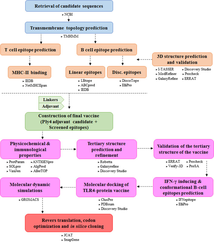
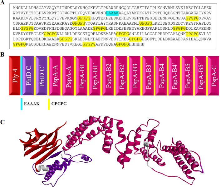
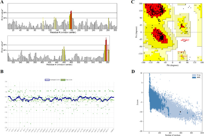
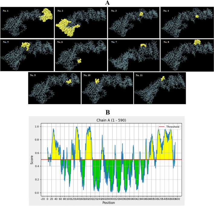
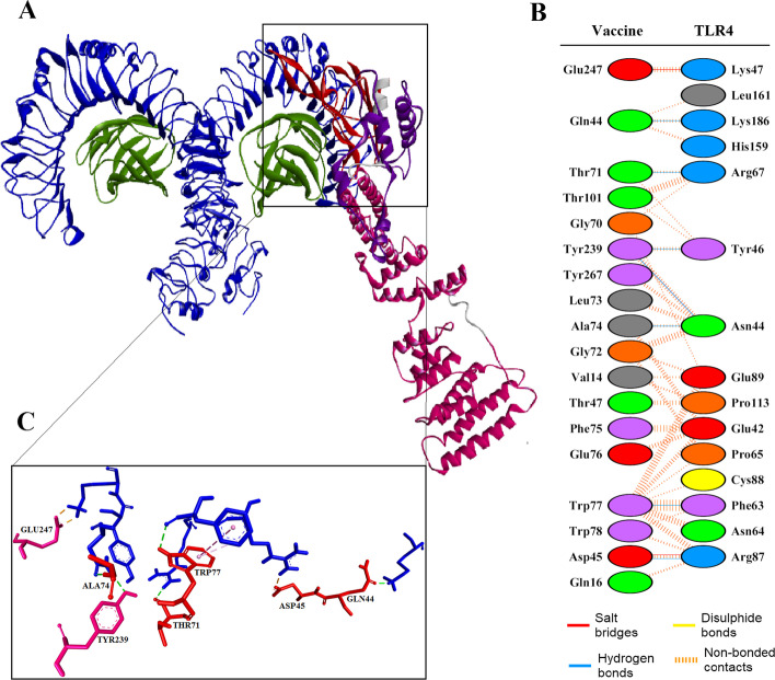
Results: The B- and T-cell epitopes from highly protective antigens PspA (clades 1-5) and PhtD were predicted and immunodominant peptides were linked to each other with proper linkers. The domain 4 of Ply, as a potential TLR4 agonist adjuvant candidate, was attached to the end of the construct to enhance the immunogenicity of the epitope vaccine. The evaluation of the physicochemical and immunological properties showed that the final construct was stable, soluble, antigenic, and non-allergenic. Furthermore, the protein was found to be acidic and hydrophilic in nature. The protein 3D-structure was built and refined, and the Ramachandran plot, ProSA-web, ERRAT, and Verify3D validated the quality of the final model. Molecular docking analysis showed that the designed construct via Ply domain 4 had a strong interaction with TLR4. The structural stability of the docked complex was confirmed by molecular dynamics. Finally, codon optimization was performed for gene expression in E. coli, followed by in silico cloning in the pET28a(+) vector.

Conclusion: The computational analysis of the construct showed acceptable results, however, the suggested vaccine needs to be experimentally verified in laboratory to ensure its safety and immunogenicity.

Keywords: Domain 4 of pneumolysin (Ply4); Immunoinformatics; Multi-epitope vaccine; Pneumococcal histidine triad protein D (PhtD); Pneumococcal surface protein A (PspA); Protein TLR agonist adjuvant.


Conflict of interest statement

The authors declare no competing interests.

Figures

Fig. 1
Fig. 1
Schematic flowchart of this study for developing the multi-epitope vaccine against S. pneumoniae. The entire approach employed in this study are represented in several phases and the used servers/softwares are mentioned below of each phase
Fig. 2
Fig. 2
The amino acid sequence, schematic diagram, and 3D refined structure of the vaccine candidate. A The final protein sequence of the multi-epitope construct consisted of 590 residues. The linkers EAAAK and GPGPG are shown in cyan and yellow, respectively. B The schematic image of vaccine construct was obtained from the selected regions of Ply4, PhtD and PspA, as well as linkers EAAAK and GPGPG, which are shown in red, purple, magenta, cyan and yellow, respectively. C The refined model was provided by the GalaxyRefine server and visualized by the Discovery Studio Visualizer. The regions of Ply4, PhtD and PspA are shown in red, purple, and magenta, respectively
Fig. 3
Fig. 3
Validation of the refined model of the multi-epitope vaccine. A The ERRAT quality factor of the refined model was estimated as 96.00. B The VERIFY-3D value of the refined model was calculated at 83.39. C Ramachandran plot of the refined model of vaccine showed 94.2%, 5.1% and 0.6% of residues in the favored, allowed, and disallowed regions, respectively. (D) Z-score of the 3D-structure of the refined model of vaccine was estimated as − 10.02, which lay in the range of scores reported for native protein structures
Fig. 4
Fig. 4
3D images of the conformational B cell epitopes of the designed vaccine and 2D score chart. A The yellow and grey regions represent the conformational epitopes and the bulk of the polyprotein, respectively. B The yellow parts with a score above the threshold 0.5 display potential B cell epitopes
Fig. 5
Fig. 5
Molecular docking of the designed vaccine with the TLR4 receptor. A The cartoon representation of the vaccine-TLR4 complex visualized by Discovery Studio Visualizer. B A number of 18 residues of the whole vaccine candidate paired with 15 residues of the TLR-4 ECD. The whole vaccine established 2 salt bridges, 7 hydrogen bonds and 116 non-bonded contacts with the receptor. C The residues involved in interactions among the vaccine and TLR4 ECD are represented as a stick model and colored in red/magenta and blue, respectively, while the vaccine residues are labeled and the interactions are shown with dashed lines
Fig. 6
Fig. 6
The results of MD simulation of the vaccine-TLR4 docked complex. A RMSD values for the vaccine-receptor complex during a period of 20 ns. The complex became stable after 6 ns of MD simulation, which further confirmed the interaction between the vaccine and TLR4. B and C show the RMSF of all the residues of construct and TLR4 ECD, respectively. Most of the receptor residues underwent small fluctuations. Two regions of the vaccine indicated relatively high fluctuations, showing relatively high flexibility
Fig. 7
Fig. 7
In silico cloning of the target sequence in pET28a(+) vector plasmid. The vaccine sequence between the restriction sites is represented in red color while the vector backbone is shown in black. The total length of the clone is 7066 bp

References

    1. Henriques-Normark B, Tuomanen EI. The pneumococcus: epidemiology, microbiology, and pathogenesis. Cold Spring Harb Perspect Med. 2013;3:a010215. doi: 10.1101/cshperspect.a010215. - DOI - PMC - PubMed
    1. Denoël P, Philipp MT, Doyle L, Martin D, Carletti G, Poolman JT. A protein-based pneumococcal vaccine protects rhesus macaques from pneumonia after experimental infection with Streptococcus pneumoniae. Vaccine. 2011;29:5495–5501. doi: 10.1016/j.vaccine.2011.05.051. - DOI - PMC - PubMed
    1. Berical AC, Harris D, Dela Cruz CS, Possick JD. Pneumococcal vaccination strategies. An update and perspective. Ann Am Thorac Soc. 2016;13:933–944. doi: 10.1513/AnnalsATS.201511-778FR. - DOI - PMC - PubMed
    1. Abdollahi S, Siadat SD, Shapouri R, Mirzaei B, Mousavi SF, Nikbin VS, Moosavi SH. Antibiotic susceptibility and prevalence of adhesion genes in Streptococcus pneumoniae isolates detected in carrier children in Tehran. Jundishapur J Microbiol. 2018;11:6. doi: 10.5812/jjm.13256. - DOI
    1. Sender V, Hentrich K, Henriques-Normark B. Virus-induced changes of the respiratory tract environment promote secondary infections with Streptococcus pneumoniae. Front Cell Infect Microbiol. 2021;11:199. doi: 10.3389/fcimb.2021.643326. - DOI - PMC - PubMed