. 2023 Jan 21;12(2):171.
doi: 10.3390/biology12020171.

Selective Destabilization of Transcripts by mRNA Decapping Regulates Oocyte Maturation and Innate Immunity Gene Expression during Ageing in C. elegans

Affiliations

Selective Destabilization of Transcripts by mRNA Decapping Regulates Oocyte Maturation and Innate Immunity Gene Expression during Ageing in C. elegans

Fivos Borbolis et al. Biology (Basel). .

Abstract

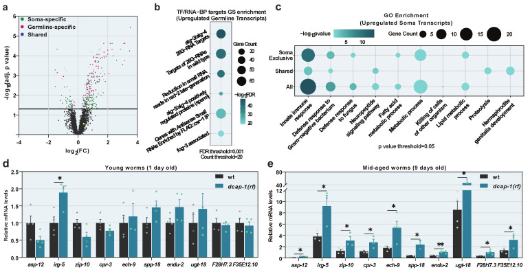
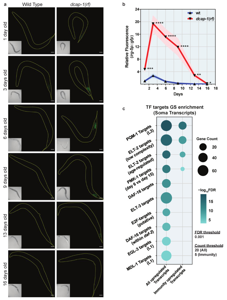
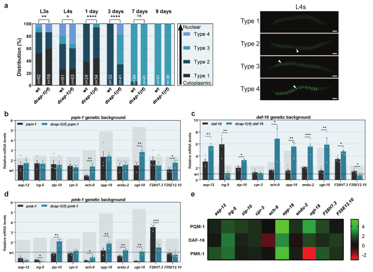
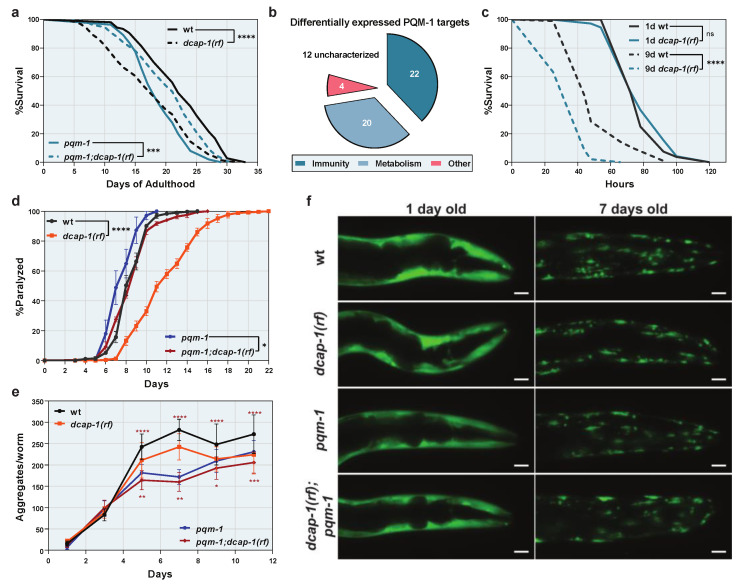
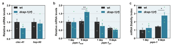
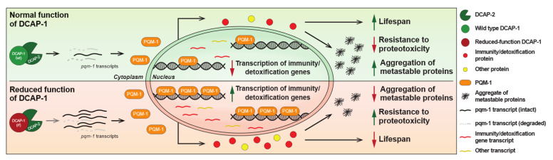
Removal of the 5' cap structure of RNAs (termed decapping) is a pivotal event in the life of cytoplasmic mRNAs mainly catalyzed by a conserved holoenzyme, composed of the catalytic subunit DCP2 and its essential cofactor DCP1. While decapping was initially considered merely a step in the general 5'-3' mRNA decay, recent data suggest a great degree of selectivity that plays an active role in the post-transcriptional control of gene expression, and regulates multiple biological functions. Studies in Caenorhabditis elegans have shown that old age is accompanied by the accumulation of decapping factors in cytoplasmic RNA granules, and loss of decapping activity shortens the lifespan. However, the link between decapping and ageing remains elusive. Here, we present a comparative microarray study that was aimed to uncover the differences in the transcriptome of mid-aged dcap-1/DCP1 mutant and wild-type nematodes. Our data indicate that DCAP-1 mediates the silencing of spermatogenic genes during late oogenesis, and suppresses the aberrant uprise of immunity gene expression during ageing. The latter is achieved by destabilizing the mRNA that encodes the transcription factor PQM-1 and impairing its nuclear translocation. Failure to exert decapping-mediated control on PQM-1 has a negative impact on the lifespan, but mitigates the toxic effects of polyglutamine expression that are involved in human disease.

Keywords: C. elegans; DCAP-1; PQM-1; ageing; innate immunity; mRNA decapping; microarray; polyglutamine; spermatogenesis.


Conflict of interest statement

The authors declare no conflict of interest.

Figures

Figure 1
Figure 1
Reduced function of DCAP-1 alters gene expression. (a) Differentially expressed genes in 9 day-old dcap-1(rf) adult worms. Gray dots correspond to non-significant expression changes (adjusted p-value > 0.05). Green dots correspond to genes expressed in the soma, magenta in the germline and blue in both. Yellow dot corresponds to pqm-1 transcript. See also Table S3. (b) Gene set (GS) enrichment analysis for transcription factor (TF) or RNA binding protein (RNA-BP) targets in differentially expressed germline-specific transcripts. (c) Gene ontology (GO) enrichment analysis in upregulated somatic transcripts. Dot size corresponds to gene count in each category. Dot color corresponds to enrichment significance. See also Table S5. (d,e) Relative mRNA levels of immunity related genes in 1 day and 9 day-old dcap-1(rf) animals, determined by qRT-PCR. Symbols represent individual values. Bars represent mean ± SEM. All values are expressed relative to 1 day-old wt animals. * p ≤ 0.05, ** p ≤ 0.01. Unpaired t-tests.
Figure 2
Figure 2
Reduced DCAP-1 function induces a transcriptional response. (a) Representative confocal images (maximum projections) of wt and dcap-1(rf) animals that express irg-5p::gfp at various ages. Scale bar = 50μm. (b) Relative fluorescence (mean ± SEM) of wt and dcap-1(rf) animals that express irg-5p::gfp at various ages. See also Figure S2. * p ≤ 0.05, ** p ≤ 0.01, *** p ≤ 0.001, **** p ≤ 0.0001. Unpaired t-test. (c) Gene set (GS) enrichment analysis for transcription factor (TF) targets in all upregulated somatic transcripts of dcap-1(rf) animals or in those related to immunity. Dot size corresponds to gene count in each category. Dot color corresponds to enrichment significance. See also Table S5.
Figure 3
Figure 3
DCAP-1 dysfunction activates PQM-1 and leads to the induction of immunity genes during ageing. (a) Distribution of wt and dcap-1(rf) worms according the degree of PQM-1 nuclear localization at various ages. n = number of worms. Images (maximum projections of confocal stacks) correspond to L4 worms, categorized depending on the degree of PQM-1 nuclear localization. White arrows point to nuclei. Scale bar = 50μm. See also Figure S3. (bd) Relative mRNA levels of immunity related genes determined by qRT-PCR in 9 day-old worms with pqm-1, daf-16 or pmk-1 mutant background. Symbols represent individual values. Bars represent mean ± SEM. All values are expressed relative to 9 day-old wt animals (dashed line). Grey rectangular backgrounds correspond to mRNA levels in single dcap-1(rf) mutants. (e) Heat map depicting the involvement of PQM-1, DAF-16 and PMK-1 in the differential expression of selected immunity-related genes in a dcap-1(rf) mutant background. Values correspond to differences in fold change (ΔFC) between single dcap-1(rf) and double pqm-1;dcap-1(rf), daf-16;dcap-1(rf) and pmk-1;dcap-1(rf) mutants, compared to their respective controls. Green shades represent a positive effect and red a negative one. * p ≤ 0.05, ** p ≤ 0.01, *** p ≤ 0.001, **** p ≤ 0.0001. Chi-square test (a), Unpaired t-tests (bd).
Figure 4
Figure 4
PQM-1 induction is detrimental for the lifespan of dcap-1(rf) animals but provides resistance to proteotoxicity. (a) Lifespan of dcap-1(rf) worms (dashed lines) in the presence or the absence of PQM-1, compared to their respective wt controls (solid lines). (b) Functional categorization of predicted PQM-1 targets, differentially expressed in dcap-1(rf) animals. See also Table S8. (c) Survival of 1 day (solid lines) and 9 day-old (dashed lines) wt and dcap-1(rf) worms after exposure to P. aeruginosa PA14. (de) Paralysis rate (d) (mean ± SEM) and number of aggregates per worm (e) (mean ± SD) of dcap-1(rf) animals that express Q35::YFP in their muscles, in the presence (orange) or the absence (red) of PQM-1, compared to their respective controls (black stars indicate comparisons between dcap-1(rf) and wt, while red stars indicate comparisons between pqm-1 and pqm-1;dcap-1(rf)). (f) Representative fluorescent images (maximum projections) showing the aggregation of Q35::YFP peptides during ageing in dcap-1(rf) animals, in the presence or absence of PQM-1, compared to their respective controls. Scale bar = 20 μm. See also Figure S4. * p < 0.05, ** p < 0.01, *** p < 0.001, **** p < 0.0001. Log-rank (Mantel-Cox) test (a,c), Two-way ANOVA (d), One-way ANOVA with Sidak’s correction (e).
Figure 5
Figure 5
PQM-1 activation is traced to the stabilization of pqm-1 transcripts in ageing dcap-1(rf) animals. (a) Relative mRNA levels of clec-41 and hsp-90 in wt and dcap-1(rf) 9 day-old worms. (b,c) Relative levels of mature (mRNAmat) and primary (mRNApri) pqm-1 transcripts (b) and stability of mature pqm-1 transcripts (c) in wt and dcap-1(rf) worms at the 1st and the 9th day of adulthood. Symbols represent individual values. Bars represent mean ± SEM. * p < 0.05, ** p < 0.01. Unpaired t-test.
Figure 6
Figure 6
Proposed model for DCAP-1-mediated regulation of longevity and resistance to proteotoxicity via PQM-1 activity. Reduced function of DCAP-1 selectively stabilizes pqm-1 mRNA transcripts, resulting in increased PQM-1 expression and its translocation to the nucleus, where it induces the expression mainly of immunity/detoxification-related genes. The resulting accrual of the corresponding proteins has a negative impact on lifespan under normal conditions, but reduces the formation of protein aggregates and favors survival under extreme proteotoxic conditions, caused by metastable peptides.

References

    1. Galloway A., Cowling V.H. mRNA cap regulation in mammalian cell function and fate. Biochim. Biophys. Acta Gene Regul. Mech. 2019;1862:270–279. doi: 10.1016/j.bbagrm.2018.09.011. - DOI - PMC - PubMed
    1. Nagarajan V.K., Jones C.I., Newbury S.F., Green P.J. XRN 5′→3′ exoribonucleases: Structure, mechanisms and functions. Biochim. Biophys. Acta. 2013;1829:590–603. doi: 10.1016/j.bbagrm.2013.03.005. - DOI - PMC - PubMed
    1. Mukherjee C., Patil D.P., Kennedy B.A., Bakthavachalu B., Bundschuh R., Schoenberg D.R. Identification of cytoplasmic capping targets reveals a role for cap homeostasis in translation and mRNA stability. Cell Rep. 2012;2:674–684. doi: 10.1016/j.celrep.2012.07.011. - DOI - PMC - PubMed
    1. Standart N., Weil D. P-Bodies: Cytosolic Droplets for Coordinated mRNA Storage. Trends Genet. 2018;34:612–626. doi: 10.1016/j.tig.2018.05.005. - DOI - PubMed
    1. Borbolis F., Syntichaki P. Biological implications of decapping: Beyond bulk mRNA decay. FEBS J. 2021;289:1457–1475. doi: 10.1111/febs.15798. - DOI - PubMed