

Overexpression of E-Cadherin Is a Favorable Prognostic Biomarker in Oral Squamous Cell Carcinoma: A Systematic Review and Meta-Analysis
- PMID: 36829516
- PMCID: PMC9953277
- DOI: 10.3390/biology12020239
Overexpression of E-Cadherin Is a Favorable Prognostic Biomarker in Oral Squamous Cell Carcinoma: A Systematic Review and Meta-Analysis
Abstract
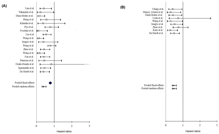
Oral squamous cell carcinoma (OSCC) is characterized by poor survival, mostly due to local invasion, loco-regional recurrence, and metastasis. Given that the weakening of cell-to-cell adhesion is a feature associated with the migration and invasion of cancer cells, different studies have explored the prognostic utility of cell adhesion molecules such as E-cadherin (E-cad). This study aims to summarize current evidence in a meta-analysis, focusing on the prognostic role of E-cad in OSCC. To find studies meeting inclusion criteria, Scopus, Web of Science, EMBASE, Medline, and OpenGrey databases were systematically assessed and screened. The selection process led to 25 studies, which were considered eligible for inclusion in the meta-analysis, representing a sample of 2553 patients. E-cad overexpression was strongly associated with longer overall survival (OS) with Hazard Ratio (HR) = 0.41 95% confidence interval (95% CI) (0.32-0.54); p < 0.001 and disease-free survival with HR 0.47 95% CI (0.37-0.61); p < 0.001. In terms of OS, patients with tongue cancer experienced better survivability when expressing E-cad with HR 0.28 95% CI (0.19-0.43); p < 0.001. Globally, our findings indicate the prognostic role of the immunohistochemical assessment of E-cad in OSCC and its expression might acquire a different role based on the oral cavity subsites.
Keywords: E-cadherin; epithelial-mesenchymal transition; meta-analysis; mouth neoplasm; prognostic; systematic review.
Conflict of interest statement
The authors declare no conflict of interest.
Figures


Similar articles
-
Prognostic impact of the loss of E-cadherin and de novo expression of N-cadherin at the invasive front of primary and recurrent oral squamous cell carcinoma.Front Oncol. 2023 May 17;13:1151879. doi: 10.3389/fonc.2023.1151879. eCollection 2023. Front Oncol. 2023. PMID: 37265789 Free PMC article.
-
The Role of E-Cadherin as a Prognostic Biomarker in Head and Neck Squamous Carcinoma: A Systematic Review and Meta-Analysis.Mol Diagn Ther. 2018 Oct;22(5):523-535. doi: 10.1007/s40291-018-0351-y. Mol Diagn Ther. 2018. PMID: 30006812
-
Prognostic value of CAIX expression in oral squamous cell carcinoma: a systematic review and meta-analysis.J Enzyme Inhib Med Chem. 2020 Dec;35(1):1258-1266. doi: 10.1080/14756366.2020.1772250. J Enzyme Inhib Med Chem. 2020. PMID: 32466707 Free PMC article.
-
P-cadherin expression reduced in squamous cell carcinoma of the oral cavity: an indicatior of poor prognosis.Cancer. 2005 Mar 1;103(5):960-9. doi: 10.1002/cncr.20858. Cancer. 2005. PMID: 15685613
-
Prognostic and Clinicopathological Significance of the Aberrant Expression of β-Catenin in Oral Squamous Cell Carcinoma: A Systematic Review and Meta-Analysis.Cancers (Basel). 2022 Jan 18;14(3):479. doi: 10.3390/cancers14030479. Cancers (Basel). 2022. PMID: 35158747 Free PMC article. Review.
Cited by
-
The Patterns of P53, E-Cadherin, β-Catenin, CXCR4 and Podoplanin Expression in Oral Squamous Cell Carcinoma Suggests a Hybrid Invasion Model: an Immunohistochemical Study on Tissue Microarrays.Head Neck Pathol. 2025 Jan 7;19(1):6. doi: 10.1007/s12105-024-01745-z. Head Neck Pathol. 2025. PMID: 39776043
-
Prognostic impact of the loss of E-cadherin and de novo expression of N-cadherin at the invasive front of primary and recurrent oral squamous cell carcinoma.Front Oncol. 2023 May 17;13:1151879. doi: 10.3389/fonc.2023.1151879. eCollection 2023. Front Oncol. 2023. PMID: 37265789 Free PMC article.
-
Biomolecules Involved in Both Metastasis and Placenta Accreta Spectrum-Does the Common Pathophysiological Pathway Exist?Cancers (Basel). 2023 May 5;15(9):2618. doi: 10.3390/cancers15092618. Cancers (Basel). 2023. PMID: 37174083 Free PMC article. Review.
-
Multi-Stage Transcriptome Analysis Revealed the Growth Mechanism of Feathers and Hair Follicles during Induction Molting by Fasting in the Late Stage of Egg Laying.Biology (Basel). 2023 Oct 19;12(10):1345. doi: 10.3390/biology12101345. Biology (Basel). 2023. PMID: 37887055 Free PMC article.
References
Publication types
LinkOut - more resources
Full Text Sources
Miscellaneous
