

Medulloblastoma: From TP53 Mutations to Molecular Classification and Liquid Biopsy
- PMID: 36829544
- PMCID: PMC9952923
- DOI: 10.3390/biology12020267
Medulloblastoma: From TP53 Mutations to Molecular Classification and Liquid Biopsy
Abstract
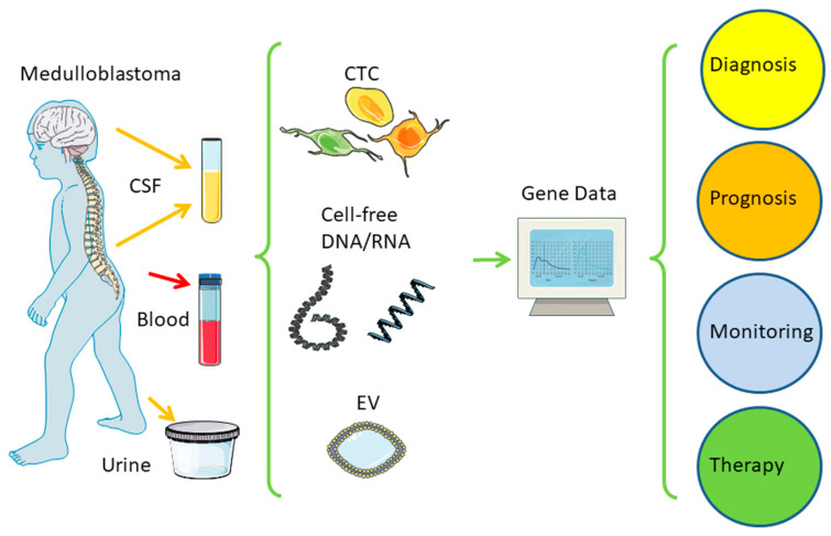
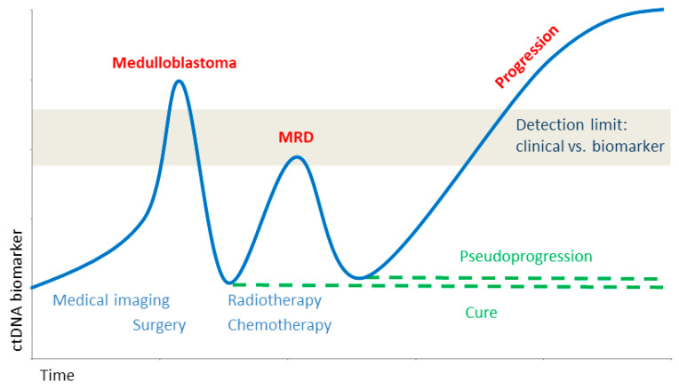
A recent paradigm shift in the diagnostics of medulloblastoma allowed the distinction of four major groups defined by genetic data rather than histology. This new molecular classification correlates better with prognosis and will allow for the better clinical management of therapies targeting druggable mutations, but also offer a new combination of monitoring tumor development in real-time and treatment response by sequential liquid biopsy. This review highlights recent developments after a century of milestones in neurosurgery and radio- and chemotherapy, but also controversial theories on the cell of origin, animal models, and the use of liquid biopsy.
Keywords: TP53 mutation; animal models; diagnostics; liquid biopsy; medulloblastoma; molecular classification; precision oncology; transcriptome.
Conflict of interest statement
The authors declare no conflict of interest.
Figures
Similar articles
-
Liquid Biopsies for Monitoring Medulloblastoma: Circulating Tumor DNA as a Biomarker for Disease Progression and Treatment Response.Cureus. 2024 Jan 5;16(1):e51712. doi: 10.7759/cureus.51712. eCollection 2024 Jan. Cureus. 2024. PMID: 38313884 Free PMC article. Review.
-
Liquid biopsy for monitoring medulloblastoma.Extracell Vesicles Circ Nucl Acids. 2022 Sep 28;3(3):280-291. doi: 10.20517/evcna.2022.36. eCollection 2022. Extracell Vesicles Circ Nucl Acids. 2022. PMID: 39697492 Free PMC article. Review.
-
Current and future applications of liquid biopsy in non-small-cell lung cancer-a narrative review.Transl Lung Cancer Res. 2023 Mar 31;12(3):594-614. doi: 10.21037/tlcr-22-742. Epub 2023 Mar 9. Transl Lung Cancer Res. 2023. PMID: 37057121 Free PMC article. Review.
-
Expression of GNAS, TP53, and PTEN Improves the Patient Prognostication in Sonic Hedgehog (SHH) Medulloblastoma Subgroup.J Mol Diagn. 2020 Jul;22(7):957-966. doi: 10.1016/j.jmoldx.2020.04.207. Epub 2020 May 4. J Mol Diagn. 2020. PMID: 32380172
-
Genetic Markers as Predictors for Response to Treatment and Possible Therapeutic Targets in Medulloblastoma.CNS Neurol Disord Drug Targets. 2023;22(5):634-642. doi: 10.2174/1871527321666220509141030. CNS Neurol Disord Drug Targets. 2023. PMID: 35579144 Review.
Cited by
-
Liquid Biopsies for Monitoring Medulloblastoma: Circulating Tumor DNA as a Biomarker for Disease Progression and Treatment Response.Cureus. 2024 Jan 5;16(1):e51712. doi: 10.7759/cureus.51712. eCollection 2024 Jan. Cureus. 2024. PMID: 38313884 Free PMC article. Review.
-
Clinical, Histological, and Molecular Prognostic Factors in Childhood Medulloblastoma: Where Do We Stand?Diagnostics (Basel). 2023 May 30;13(11):1915. doi: 10.3390/diagnostics13111915. Diagnostics (Basel). 2023. PMID: 37296767 Free PMC article. Review.
-
Liquid biopsy and glioblastoma.Explor Target Antitumor Ther. 2023;4(1):28-41. doi: 10.37349/etat.2023.00121. Epub 2023 Feb 25. Explor Target Antitumor Ther. 2023. PMID: 36937320 Free PMC article. Review.
-
Integrative Analysis of the Role of TP53 in Human Pan-Cancer.Curr Issues Mol Biol. 2023 Nov 29;45(12):9606-9633. doi: 10.3390/cimb45120601. Curr Issues Mol Biol. 2023. PMID: 38132447 Free PMC article.
References
-
- Ohgaki H., Eibl R.H., Wiestler O.D., Yasargil M.G., Newcomb E.W., Kleihues P. P53 Mutations in Nonastrocytic Human Brain Tumors. Cancer Res. 1991;51:6202–6205. - PubMed
-
- Louis D.N., Perry A., Reifenberger G., von Deimling A., Figarella-Branger D., Cavenee W.K., Ohgaki H., Wiestler O.D., Kleihues P., Ellison D.W. The 2016 World Health Organization Classification of Tumors of the Central Nervous System: A Summary. Acta Neuropathol. 2016;131:803–820. doi: 10.1007/s00401-016-1545-1. - DOI - PubMed
-
- Louis D.N., Perry A., Wesseling P., Brat D.J., Cree I.A., Figarella-Branger D., Hawkins C., Ng H.K., Pfister S.M., Reifenberger G., et al. The 2021 WHO Classification of Tumors of the Central Nervous System: A Summary. Neuro-Oncology. 2021;23:1231–1251. doi: 10.1093/neuonc/noab106. - DOI - PMC - PubMed
-
- Bailey P., Cushing H. Medulloblastoma Cerebelli: A Common Type of Midcerebellar Glioma of the Childhood. Arch. Neurol. Psychiatry. 1925;14:192–224. doi: 10.1001/archneurpsyc.1925.02200140055002. - DOI
Publication types
LinkOut - more resources
Full Text Sources
Research Materials
Miscellaneous
