. 2023 Feb 16;12(2):318.
doi: 10.3390/biology12020318.

Reduced Renal CSE/CBS/H2S Contributes to the Progress of Lupus Nephritis

Affiliations

Reduced Renal CSE/CBS/H2S Contributes to the Progress of Lupus Nephritis

Xuan Wang et al. Biology (Basel). .

Abstract

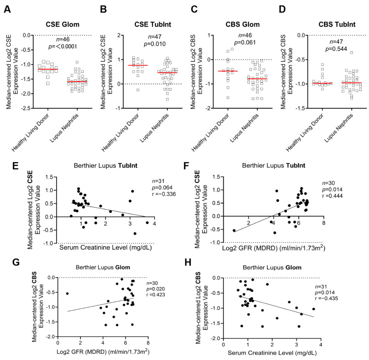
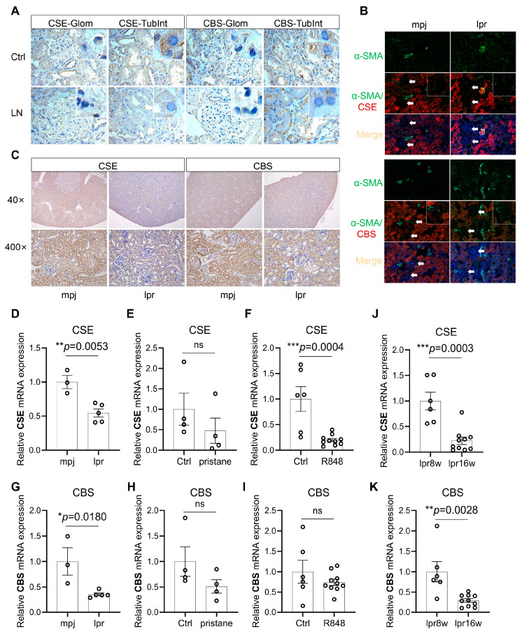
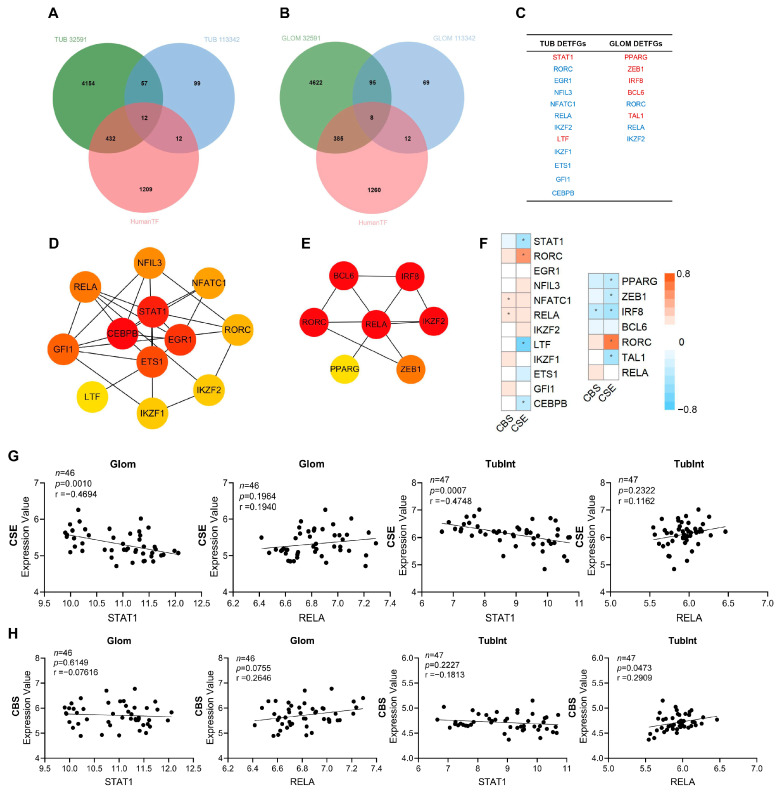
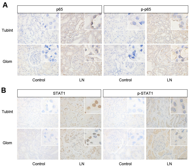
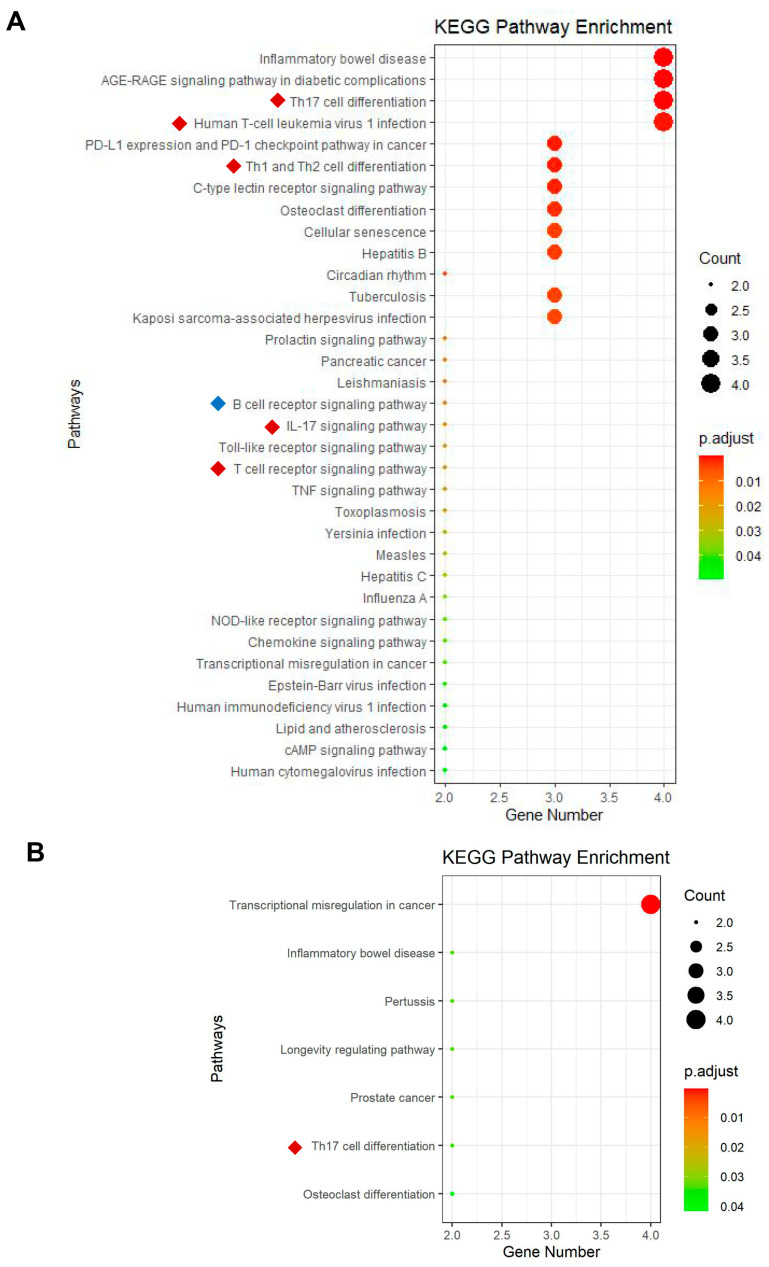
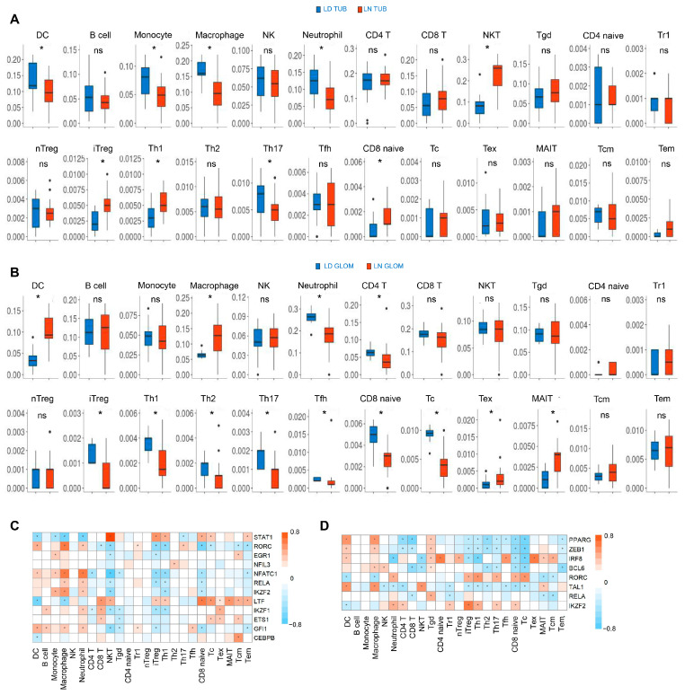
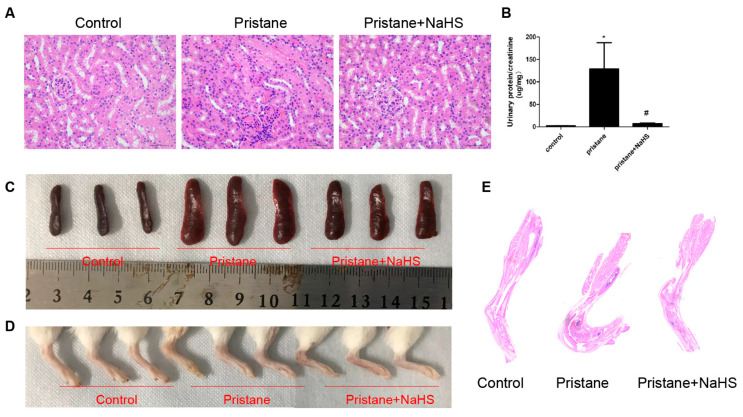
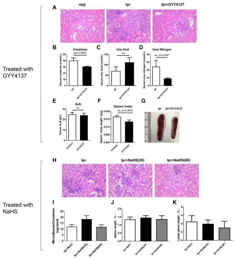
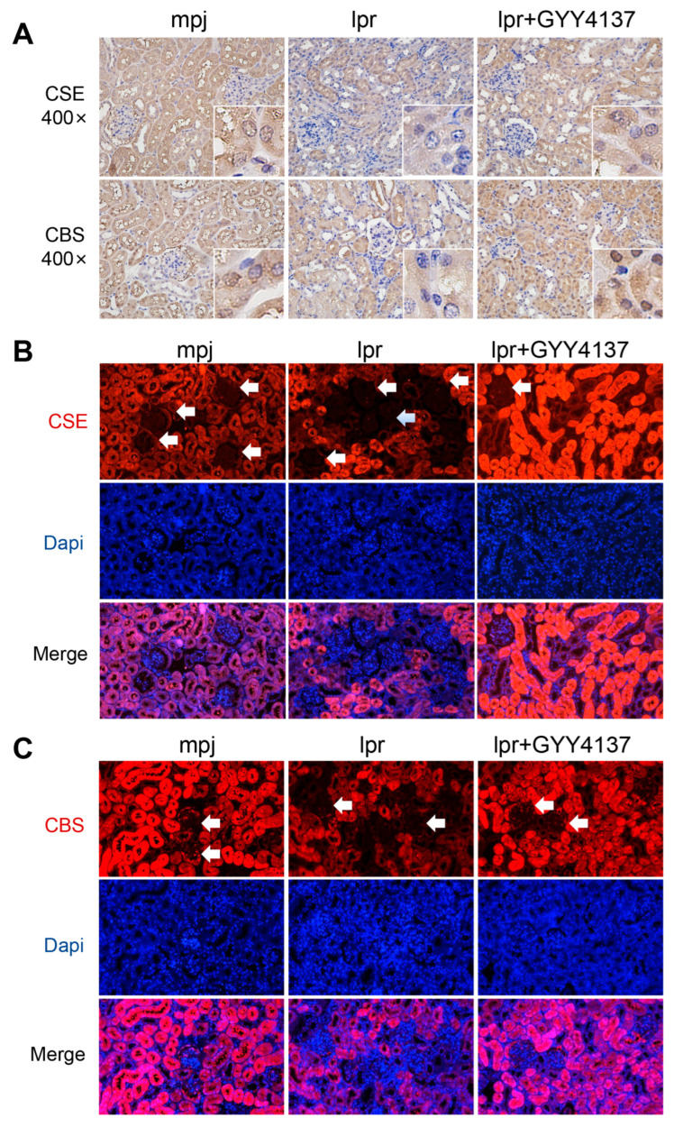
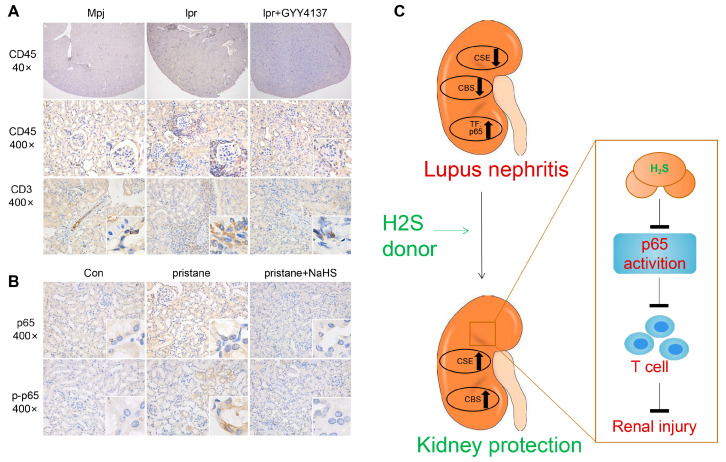
The molecular mechanisms underlying lupus nephritis (LN) pathogenesis are not fully understood. Hydrogen sulfide (H2S) is involved in many pathological and physiological processes. We sought to investigate the roles of H2S in LN pathogenesis. H2S synthase cystathionine-lyase (CSE) and cystathionine-synthetase (CBS) expression was downregulated in renal tissues of patients with LN and their levels were associated with LN's prognosis using the Nephroseq database. Reduced CSE and CBS protein expression in kidney tissues of LN patients and MRL/lpr mice were confirmed by immunohistochemistry. CSE and CBS mRNA levels were reduced in MRL/lpr and pristine- and R848-induced lupus mice. Given that H2S exerts an anti-inflammatory role partly via regulating inflammatory transcription factors (TFs), we analyzed hub TFs by using a bioinformatics approach. It showed that STAT1, RELA, and T-cell-related signaling pathways were enriched in LN. Increased STAT1 and RELA expression were confirmed in renal tissues of LN patients. Treatment of MRL/lpr and pristine mice with H2S donors alleviated systemic lupus erythematosus (SLE) phenotypes and renal injury. H2S donors inhibited RELA level and T-cell infiltration in the kidneys of MRL/lpr and pristine mice. Our data indicated that CSE/CBS/H2S contributes to LN pathogenesis. Supplementation of H2S would be a potential therapeutic strategy for LN.

Keywords: CBS; CSE; H2S; RELA; STAT1; T cells; lupus nephritis.


Conflict of interest statement

The authors declare no conflict of interest.

Figures

Figure 1
Figure 1
Downregulation of hydrogen sulfide synthetase in the kidneys of lupus nephritis patients was associated with poor renal prognosis. (A,B) Gene expression of renal CSE in Glom and TubInt of healthy living donors and LN patients (Nephroseq database). (C,D) Gene expression of renal CBS in Glom and TubInt of healthy living donors and LN patients. (E,F) Correlation of renal TubInt CSE gene expression with serum creatinine and GFR in patients with LN in Nephroseq database. (G,H) Correlation of renal Glom CBS gene expression with serum creatinine and GFR in patients with LN in Nephroseq database.
Figure 2
Figure 2
CSE and CBS were decreased in the kidney tissue of lupus mice. (A) Representative immunohistochemical staining images of CSE and CBS from human renal-puncture tissue (400×). Glom: glomeruli; TubInt: tubulointerstitium; n: total number of data; Ctrl: normal or minimal change kidney of the donor. (B) Representative immunofluorescence staining images of CSE, CBS, DAPI, and αSMA (a marker of glomerular mesangial cells and renal interstitial vascular smooth muscle cells) from the kidney of MRL/mpj and MRL/lpr mice (200×); the white arrows and enlarge box in the upper right corner of pictures refer to the glomerulus. (C) Representative immunohistochemical staining images of CSE and CBS from the kidney of MRL/mpj and MRL/lpr mice (40× and 400×). (D) mRNA expression of CSE in 16-week-old MRL/mpj and MRL/lpr mice. (E) mRNA expression of CSE in control Balb/c and pristane-induced Balb/c mice. (F) mRNA expression of CSE in control Balb/c and R848-induced Balb/c mice. (G) mRNA expression of CBS in 16-week-old MRL/mpj and MRL/lpr mice. (H) mRNA expression of CBS in control Balb/c and pristane-induced Balb/c mice. (I) mRNA expression of CBS in control Balb/c and R848-induced Balb/c mice. (J,K) mRNA expression of CSE and CBS in 8-week-old (pre-diseased) and 16-week-old (severely diseased) MRL/lpr mice. *, p < 0.05; **, p < 0.01; ***, p < 0.001.
Figure 3
Figure 3
Screen of key TFs in renal tissue of patients with LN and its correlation with CBS and CSE. The Venn diagram of the differential TFs in the tubulointerstitium (A) and glomeruli (B), based on GSE32591, GSE113342, and human TFDB. (C) Red represents upregulation and blue represents downregulation for the differentially expressed TF genes (DETFGs) in the tubulointerstitium and glomeruli. (D) The interaction of hub transcription factors in the tubulointerstitium: the redder the color, the higher the degree through modules cytohHubba for sorting top hub genes. (E) The interaction of hub transcription factors in the glomeruli. (FH) Correlation analysis between gene expression of CSE, CBS, and DETFGs. TUB/TubInt, tubulointerstitium; GLOM, glomeruli; *, p < 0.05.
Figure 4
Figure 4
The expression of p65 and STAT1 in kidney tissue of LN patients. (A,B) Representative images of p65, p-p65, STAT1, and p-STAT1 staining in renal tissues of control and patients with LN (400×). TubInt, tubulointerstitium; Glom, glomeruli.
Figure 5
Figure 5
KEGG enrichment analysis of DETFGs in the tubulointerstitial and glomeruli of renal tissue of LN. (A) KEGG enrichment analysis of DETFGs in the tubulointerstitium; red and blue markers represent the signaling pathways associated with T cells and B cells, respectively. (B) KEGG enrichment analysis of DETFGs in glomeruli.
Figure 6
Figure 6
Immune cell subtype infiltration in the tubulointerstitial and glomeruli of renal tissue of LN. (A,B) The proportion of the 26 types of immune cell infiltration in the tubulointerstitium (A) and glomeruli (B). (C,D) Association between TFs and immune cell populations in LN. The association of TFs with immune cell populations in the tubulointerstitium (C) and glomeruli (D). Tgd, gamma delta T cells; Tr1, type 1 regulatory T cells; nTreg, natural T-regulatory cells; Tcm, central memory T; *, p < 0.05.
Figure 7
Figure 7
NaHS-attenuated pristane-induced renal injury, splenic proliferation, and arthritis. (A) Representative images of mouse renal tissues after HE staining (400×). (B) The ratio of urine protein and urine creatinine from mice was measured. Data are represented as the means ± SEM (n = 5). * p < 0.01, vs. control group. # p < 0.01, vs. pristane-induced lupus group. (C,D) Images of spleens and the lower ankle from control, pristane-induced with or without NaHS mice are shown. (E) Representative scan images of mouse lower ankle after HE staining.
Figure 8
Figure 8
The H2S donors, GYY4137 and NaHS, partially attenuated renal injury in MRL/lpr mice. (A) Representative images of mouse renal tissues after HE staining in GYY4137-treated mice (400×). (BE) The serum levels of creatinine, urea nitrogen, uric acid, and albumin were detected upon GYY4137 injection. (F,G) The ratio of spleen weight and body weight and representative images of the spleen have been shown. Data are represented as the means ± SEM (n = 6). * p < 0.05. (H) Representative images of mouse renal tissues after HE staining in NaHS-treated mice. (I) The ratio of urine protein and urine creatinine from mice was measured. (J,K) The ratio of spleen or cervical lymph node weight and body weight was calculated.
Figure 9
Figure 9
H2S donor recovers the reduced expression of CSE and CBS in MRL/lpr mice. (A) Representative images of mouse renal tissues after IHC staining of CSE and CBS in MRL/lpr and MRL/mpj mice (400×). (B,C) Representative images of mouse renal tissues after IF staining of CSE, CBS, and DAPI in MRL/lpr and MRL/mpj mice (200×); the white arrow refers to the glomerulus.
Figure 10
Figure 10
H2S supplementation reduces the expression of p65 and T-cell infiltration in the kidney of lupus mice. (A,B) Representative images of mouse renal tissues after IHC staining of p65 and p-p65 in pristane-induced mice (400×). (C) A model depicting the potential role of the H2S donor in the attenuation of LN.

Similar articles

Cited by

References

    1. Yu F., Haas M., Glassock R., Zhao M.-H. Redefining lupus nephritis: Clinical implications of pathophysiologic subtypes. Nat. Rev. Nephrol. 2017;13:483–495. doi: 10.1038/nrneph.2017.85. - DOI - PubMed
    1. Yu C., Li P., Dang X., Zhang X., Mao Y., Chen X. Lupus nephritis: New progress in diagnosis and treatment. J. Autoimmun. 2022;132:102871. doi: 10.1016/j.jaut.2022.102871. - DOI - PubMed
    1. Almaani S., Meara A., Rovin B.H. Update on Lupus Nephritis. Clin. J. Am. Soc. Nephrol. 2017;12:825–835. doi: 10.2215/CJN.05780616. - DOI - PMC - PubMed
    1. Aziz F., Chaudhary K. Lupus Nephritis: A Treatment Update. Curr. Clin. Pharmacol. 2018;13:4–13. doi: 10.2174/1574884713666180403150359. - DOI - PubMed
    1. Davidson A., Aranow C., Mackay M. Lupus nephritis: Challenges and progress. Curr. Opin. Rheumatol. 2019;31:682–688. doi: 10.1097/BOR.0000000000000642. - DOI - PMC - PubMed