

Deep Learning-Based Medical Images Segmentation of Musculoskeletal Anatomical Structures: A Survey of Bottlenecks and Strategies
- PMID: 36829631
- PMCID: PMC9952222
- DOI: 10.3390/bioengineering10020137
Deep Learning-Based Medical Images Segmentation of Musculoskeletal Anatomical Structures: A Survey of Bottlenecks and Strategies
Abstract
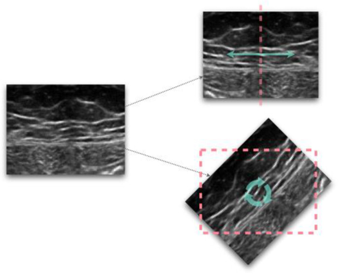
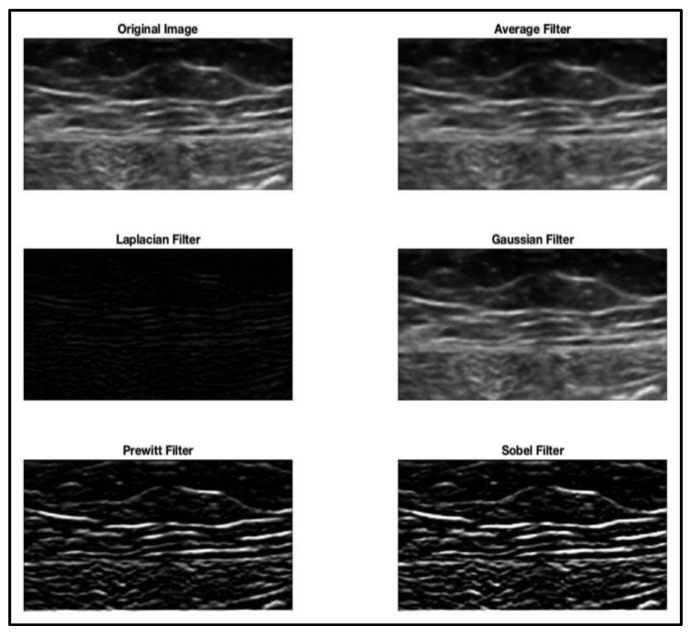
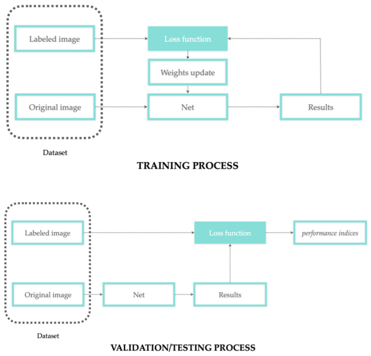
By leveraging the recent development of artificial intelligence algorithms, several medical sectors have benefited from using automatic segmentation tools from bioimaging to segment anatomical structures. Segmentation of the musculoskeletal system is key for studying alterations in anatomical tissue and supporting medical interventions. The clinical use of such tools requires an understanding of the proper method for interpreting data and evaluating their performance. The current systematic review aims to present the common bottlenecks for musculoskeletal structures analysis (e.g., small sample size, data inhomogeneity) and the related strategies utilized by different authors. A search was performed using the PUBMED database with the following keywords: deep learning, musculoskeletal system, segmentation. A total of 140 articles published up until February 2022 were obtained and analyzed according to the PRISMA framework in terms of anatomical structures, bioimaging techniques, pre/post-processing operations, training/validation/testing subset creation, network architecture, loss functions, performance indicators and so on. Several common trends emerged from this survey; however, the different methods need to be compared and discussed based on each specific case study (anatomical region, medical imaging acquisition setting, study population, etc.). These findings can be used to guide clinicians (as end users) to better understand the potential benefits and limitations of these tools.
Keywords: CT; MRI; X-ray; anatomical structures; artificial intelligence; deep learning; medical imaging; musculoskeletal system; segmentation; ultrasonography.
Conflict of interest statement
The funders had no role in the design of the study; in the collection, analyses, or interpretation of data; in the writing of the manuscript; or in the decision to publish the results.
Figures
References
-
- Kompella G., Antico M., Sasazawa F., Jeevakala S., Ram K., Fontanarosa D., Pandey A.K., Sivaprakasam M. Segmentation of Femoral Cartilage from Knee Ultrasound Images Using Mask R-CNN. Proc. Annu. Int. Conf. IEEE Eng. Med. Biol. Soc. EMBS. 2019;2019:966–969. doi: 10.1109/EMBC.2019.8857645. - DOI - PubMed
Publication types
Grants and funding
LinkOut - more resources
Full Text Sources
