

Exfoliative Cytology and Genetic Analysis for a Non-Invasive Approach to the Diagnosis of White Sponge Nevus: Case Series
- PMID: 36829648
- PMCID: PMC9952746
- DOI: 10.3390/bioengineering10020154
Exfoliative Cytology and Genetic Analysis for a Non-Invasive Approach to the Diagnosis of White Sponge Nevus: Case Series
Abstract
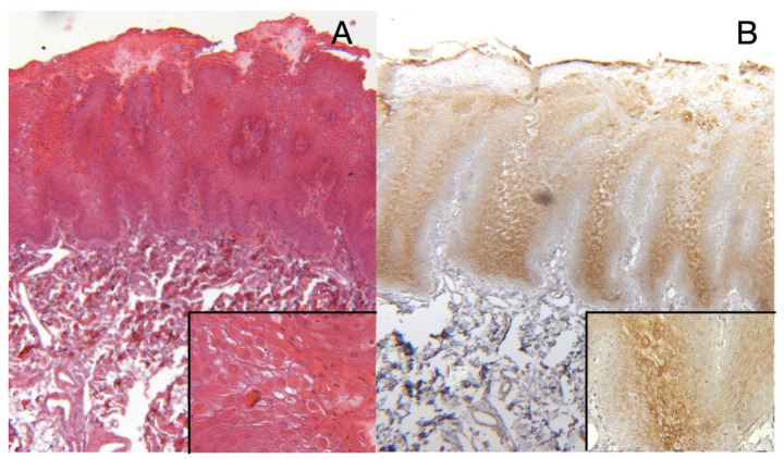
Background: White Sponge Nevus (WSN) is a rare benign disorder associated with mutations in genes coding for cytokeratin 4 (KRT4) and 13 (KRT13) characterized by dyskeratotic hyperplasia of mucous membranes. This study was aimed at examining different approaches (cytology, pathology and genetic analysis) to WSN diagnosis.
Methods: A series of four patients with asymptomatic white diffuse oral lesions were evaluated and, before performing an incisional biopsy for pathology, an oral brush Thin Prep was collected for exfoliative liquid-based cytology (LBC). DNA for genetic analysis was also obtained from patients and both their parents, using buccal swabs.
Results: Pathology and cytology showed similar results, leading to the same diagnosis of hyperkeratotic epithelium with acanthosis and spongiosis, without atypia, demonstrating the efficiency of LBC for the differential diagnosis. Sequencing analysis revealed at least 6 rare variants in the KRT4 and KRT13 genes in each patient, contributed in part by both unaffected parents.
Conclusions: Thin Prep for oral exfoliative cytology and genetic analysis are sufficient for an accurate diagnosis of WSN. The combination of cytological and genetic analyses could substitute the histologic exam, providing a non-invasive alternative for incisional biopsy.
Keywords: Cell Block; KRT13; KRT4; White Sponge Nevus (WSN); incisional biopsy; liquid-based cytology.
Conflict of interest statement
This research did not receive any specific grant from funding agencies in the public, commercial, or not-for-profit sectors.
Figures


Similar articles
-
Clinical features and molecular genetic analysis in a Turkish family with oral white sponge nevus.Med Oral Patol Oral Cir Bucal. 2018 Mar 1;23(2):e144-e150. doi: 10.4317/medoral.21437. Med Oral Patol Oral Cir Bucal. 2018. PMID: 29476668 Free PMC article.
-
Keratin 13 mutations associated with oral white sponge nevus in two Chinese families.Meta Gene. 2014 May 17;2:374-83. doi: 10.1016/j.mgene.2014.04.008. eCollection 2014 Dec. Meta Gene. 2014. PMID: 25606422 Free PMC article.
-
Current approaches to the diagnosis and treatment of white sponge nevus.Expert Rev Mol Med. 2015 May 29;17:e9. doi: 10.1017/erm.2015.7. Expert Rev Mol Med. 2015. PMID: 26021387 Review.
-
A novel mutation in the keratin 4 gene causing white sponge naevus.Br J Dermatol. 2003 Jun;148(6):1125-8. doi: 10.1046/j.1365-2133.2003.05337.x. Br J Dermatol. 2003. PMID: 12828738
-
White sponge nevus presenting in the esophagus--case report and literature review.Pathology. 1992 Apr;24(2):112-5. doi: 10.3109/00313029209063635. Pathology. 1992. PMID: 1641256 Review.
References
LinkOut - more resources
Full Text Sources
Research Materials
Miscellaneous
