. 2023 Feb 1;10(2):181.
doi: 10.3390/bioengineering10020181.

Comparing 3D, 2.5D, and 2D Approaches to Brain Image Auto-Segmentation

Affiliations

Comparing 3D, 2.5D, and 2D Approaches to Brain Image Auto-Segmentation

Arman Avesta et al. Bioengineering (Basel). .

Abstract

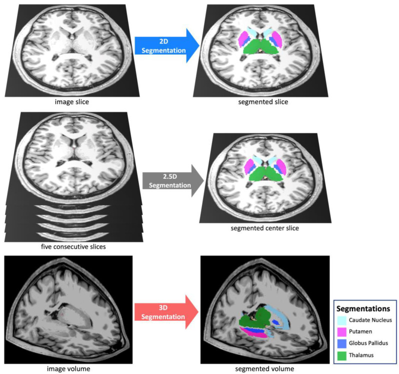
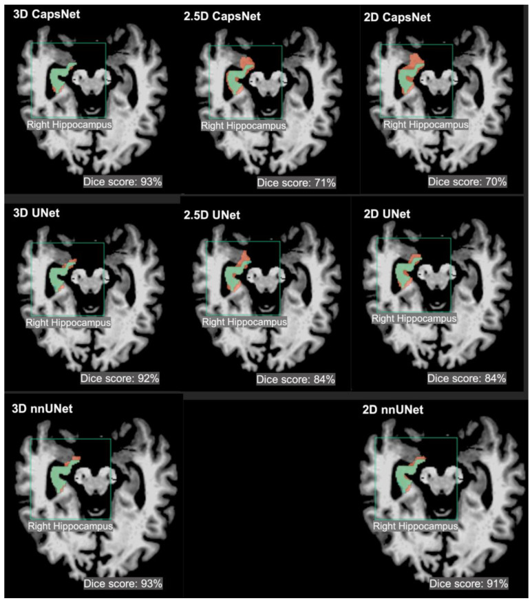
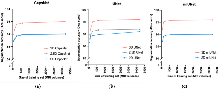
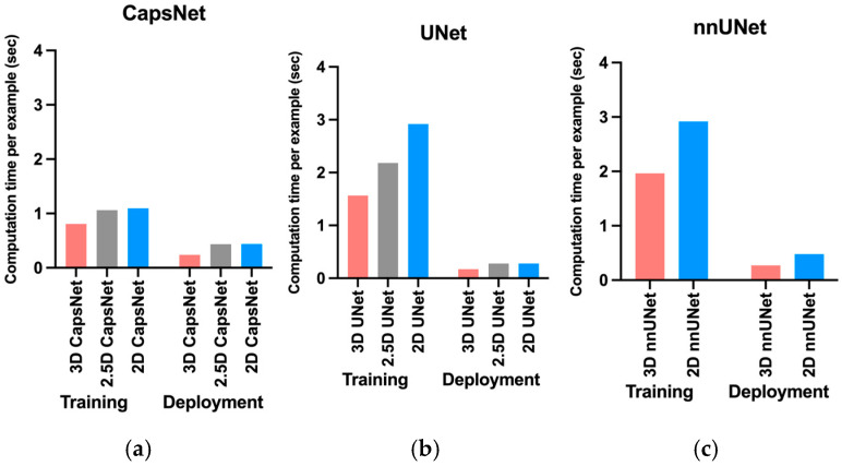
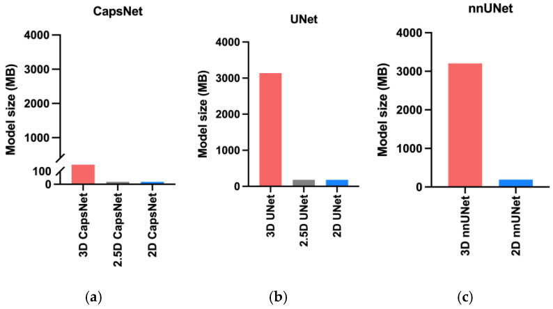
Deep-learning methods for auto-segmenting brain images either segment one slice of the image (2D), five consecutive slices of the image (2.5D), or an entire volume of the image (3D). Whether one approach is superior for auto-segmenting brain images is not known. We compared these three approaches (3D, 2.5D, and 2D) across three auto-segmentation models (capsule networks, UNets, and nnUNets) to segment brain structures. We used 3430 brain MRIs, acquired in a multi-institutional study, to train and test our models. We used the following performance metrics: segmentation accuracy, performance with limited training data, required computational memory, and computational speed during training and deployment. The 3D, 2.5D, and 2D approaches respectively gave the highest to lowest Dice scores across all models. 3D models maintained higher Dice scores when the training set size was decreased from 3199 MRIs down to 60 MRIs. 3D models converged 20% to 40% faster during training and were 30% to 50% faster during deployment. However, 3D models require 20 times more computational memory compared to 2.5D or 2D models. This study showed that 3D models are more accurate, maintain better performance with limited training data, and are faster to train and deploy. However, 3D models require more computational memory compared to 2.5D or 2D models.

Keywords: auto-segmentation; deep learning; magnetic resonance imaging; neuroimaging.


Conflict of interest statement

The funders had no role in the design of the study; in the collection, analyses, or interpretation of data; in the writing of the manuscript; or in the decision to publish the results.

Figures

Figure 1
Figure 1
We compared three segmentation approaches: 3D, 2.5D, and 2D. The 2D approach analyzes and segments one slice of the image, the 2.5D approach analyzes five consecutive slices of the image to segment the middle slice, and the 3D approach analyzes and segments a 3D volume of the image.
Figure 2
Figure 2
Examples of 3D, 2.5D, and 2D segmentations of the right hippocampus by CapsNet, UNet, and nnUNet. Target segmentations and model predictions are respectively shown in green and red. Dice scores are provided for the entire volume of the right hippocampus in this patient (who was randomly chosen from the test set).
Figure 3
Figure 3
Comparing 3D, 2.5D, and 2D approaches when training data is limited. As we decreased the size of the training set from 3000 MRIs down to 60 MRIs, the CapsNet (a), UNet (b), and nnUNet (c) models maintained higher segmentation accuracy (measured by Dice scores).
Figure 4
Figure 4
Comparing computational time required by 3D, 2.5D, and 2D approaches to train and deploy auto-segmentation models. The training times represent how much time it would take per training example per epoch for the model to converge. The deployment times represent how much time each model would require to segment one brain MRI volume. The 3D approach trained and deployed faster across all auto-segmentation models, including CapNet (a), UNet (b), and nnUNet (c).
Figure 5
Figure 5
Comparing the memory required by the 3D, 2.5D, and 2D approaches. The bars represent the computational memory required to accommodate the total size of each model, including the parameters plus the cumulative size of the forward- and backward-pass feature volumes. Within each auto-segmentation model including the CapsNet (a), UNet (b), and nnUNet (c), the 3D approach required 20 times more computational memory compared to the 2.5D or 2D approaches.

References

    1. Feng C.H., Cornell M., Moore K.L., Karunamuni R., Seibert T.M. Automated contouring and planning pipeline for hippocampal-avoidant whole-brain radiotherapy. Radiat. Oncol. 2020;15:251. doi: 10.1186/s13014-020-01689-y. - DOI - PMC - PubMed
    1. Dasenbrock H.H., See A.P., Smalley R.J., Bi W.L., Dolati P., Frerichs K.U., Golby A.J., Chiocca E.A., Aziz-Sultan M.A. Frameless Stereotactic Navigation during Insular Glioma Resection using Fusion of Three-Dimensional Rotational Angiography and Magnetic Resonance Imaging. World Neurosurg. 2019;126:322–330. doi: 10.1016/j.wneu.2019.03.096. - DOI - PubMed
    1. Dolati P., Gokoglu A., Eichberg D., Zamani A., Golby A., Al-Mefty O. Multimodal navigated skull base tumor resection using image-based vascular and cranial nerve segmentation: A prospective pilot study. Surg. Neurol. Int. 2015;6:172. doi: 10.4103/2152-7806.170023. - DOI - PMC - PubMed
    1. Clerx L., Gronenschild H.B.M., Echavarri C., Aalten P., Jacobs H.I.L. Can FreeSurfer Compete with Manual Volumetric Measurements in Alzheimer’s Disease? Curr. Alzheimer Res. 2015;12:358–367. doi: 10.2174/1567205012666150324174813. - DOI - PubMed
    1. Bousabarah K., Ruge M., Brand J.-S., Hoevels M., Rueß D., Borggrefe J., Hokamp N.G., Visser-Vandewalle V., Maintz D., Treuer H., et al. Deep convolutional neural networks for automated segmentation of brain metastases trained on clinical data. Radiat. Oncol. 2020;15:87. doi: 10.1186/s13014-020-01514-6. - DOI - PMC - PubMed

LinkOut - more resources