. 2023 Feb 14;10(2):250.
doi: 10.3390/bioengineering10020250.

Synthetic CT in Carbon Ion Radiotherapy of the Abdominal Site

Affiliations

Synthetic CT in Carbon Ion Radiotherapy of the Abdominal Site

Giovanni Parrella et al. Bioengineering (Basel). .

Abstract

The generation of synthetic CT for carbon ion radiotherapy (CIRT) applications is challenging, since high accuracy is required in treatment planning and delivery, especially in an anatomical site as complex as the abdomen. Thirty-nine abdominal MRI-CT volume pairs were collected and a three-channel cGAN (accounting for air, bones, soft tissues) was used to generate sCTs. The network was tested on five held-out MRI volumes for two scenarios: (i) a CT-based segmentation of the MRI channels, to assess the quality of sCTs and (ii) an MRI manual segmentation, to simulate an MRI-only treatment scenario. The sCTs were evaluated by means of similarity metrics (e.g., mean absolute error, MAE) and geometrical criteria (e.g., dice coefficient). Recalculated CIRT plans were evaluated through dose volume histogram, gamma analysis and range shift analysis. The CT-based test set presented optimal MAE on bones (86.03 ± 10.76 HU), soft tissues (55.39 ± 3.41 HU) and air (54.42 ± 11.48 HU). Higher values were obtained from the MRI-only test set (MAEBONE = 154.87 ± 22.90 HU). The global gamma pass rate reached 94.88 ± 4.9% with 3%/3 mm, while the range shift reached a median (IQR) of 0.98 (3.64) mm. The three-channel cGAN can generate acceptable abdominal sCTs and allow for CIRT dose recalculations comparable to the clinical plans.

Keywords: MRI guidance; MRI-only; carbon ion radiotherapy; deep learning; image-guided radiotherapy; particle therapy; synthetic CT.


Conflict of interest statement

The authors declare no conflict of interest.

Figures
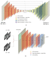
Figure 1
Figure 1
(a) U-net generator. (b) PatchGAN discriminator.
Figure 2
Figure 2
(a) Example of MRI, CT, sCT and pseudo ground truth (CTPGT) on axial plane. CTPGT still shows visible discrepancies with MRI anatomical condition. (b) Example of inter-acquisition motion in a CT-MRI pair and the resulted sCT in CV. (c) Example of MRI, planning CT and synthetic CT from MRI-only scenario. In red, the segmentation of kidneys used for the geometrical analysis.
Figure 3
Figure 3
(a) DVH comparison on patients P17 and P27; (b) original CIRT plan (RBE) and sCT-based recalculation for patients P17 and P27.
Figure 4
Figure 4
(a) D95% values for GTV and CTV in the original plan (CT) and the recalculated one (sCT). For P17, PTV was considered in this comparison (shown in Green). The table contains the dose values and the dose difference Δ[Gy[RBE]], as well as the error relative to the prescribed dose E [%]. (b) D2% difference (sCT-CT) for the main OARs on each patient, and the D2% MAE over each OAR. The red mark indicates the median, and edges of the box show the 25th and 75th percentiles.
Figure 5
Figure 5
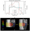
Representative range shift analysis on patient P27. (a) The graph shows both dose and HU profiles in CT (light blue) and sCT (red), evaluated along the yellow line shown in (b). The 80% reference is marked by the horizontal line. (b) Corresponding CT and sCT sagittal views are compared. The yellow line is one of the 10 considered for each beam, on different transversal slices.

References

    1. Durante M., Orecchia R., Loeffler J.S. Charged-Particle Therapy in Cancer: Clinical Uses and Future Perspectives. Nat. Rev. Clin. Oncol. 2017;14:483–495. doi: 10.1038/nrclinonc.2017.30. - DOI - PubMed
    1. Liermann J., Shinoto M., Syed M., Debus J., Herfarth K., Naumann P. Carbon Ion Radiotherapy in Pancreatic Cancer: A Review of Clinical Data. Radiother. Oncol. 2020;147:145–150. doi: 10.1016/j.radonc.2020.05.012. - DOI - PubMed
    1. Li H., Dong L., Bert C., Chang J., Flampouri S., Jee K., Lin L., Moyers M., Mori S., Rottmann J., et al. AAPM Task Group Report 290: Respiratory Motion Management for Particle Therapy. Med. Phys. 2022;49:e50–e81. doi: 10.1002/mp.15470. - DOI - PMC - PubMed
    1. Jaffray D.A. Image-Guided Radiotherapy: From Current Concept to Future Perspectives. Nat. Rev. Clin. Oncol. 2012;9:688–699. doi: 10.1038/nrclinonc.2012.194. - DOI - PubMed
    1. Otazo R., Lambin P., Pignol J.P., Ladd M.E., Schlemmer H.P., Baumann M., Hricak H. MRI-Guided Radiation Therapy: An Emerging Paradigm in Adaptive Radiation Oncology. Radiology. 2021;298:248–260. doi: 10.1148/radiol.2020202747. - DOI - PMC - PubMed

LinkOut - more resources