

Functional Loss of Terminal Complement Complex Protects Rabbits from Injury-Induced Osteoarthritis on Structural and Cellular Level
- PMID: 36830586
- PMCID: PMC9953363
- DOI: 10.3390/biom13020216
Functional Loss of Terminal Complement Complex Protects Rabbits from Injury-Induced Osteoarthritis on Structural and Cellular Level
Abstract
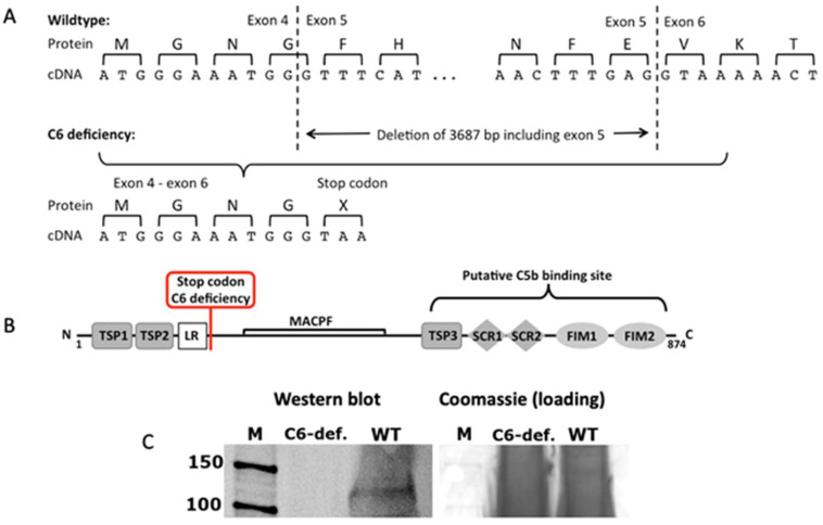
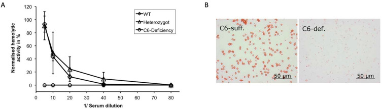
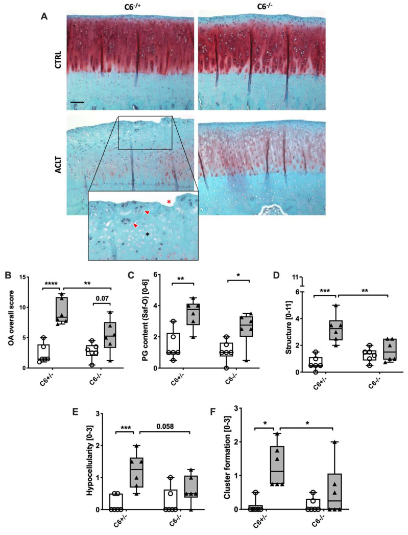
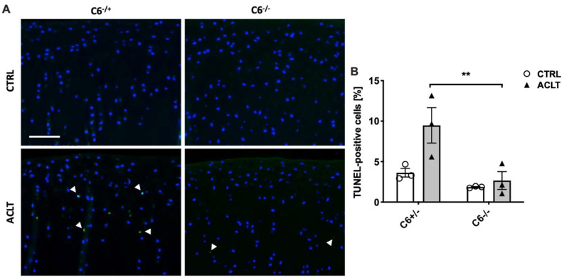
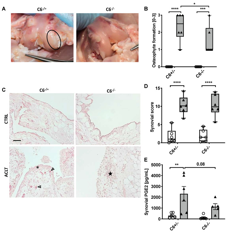
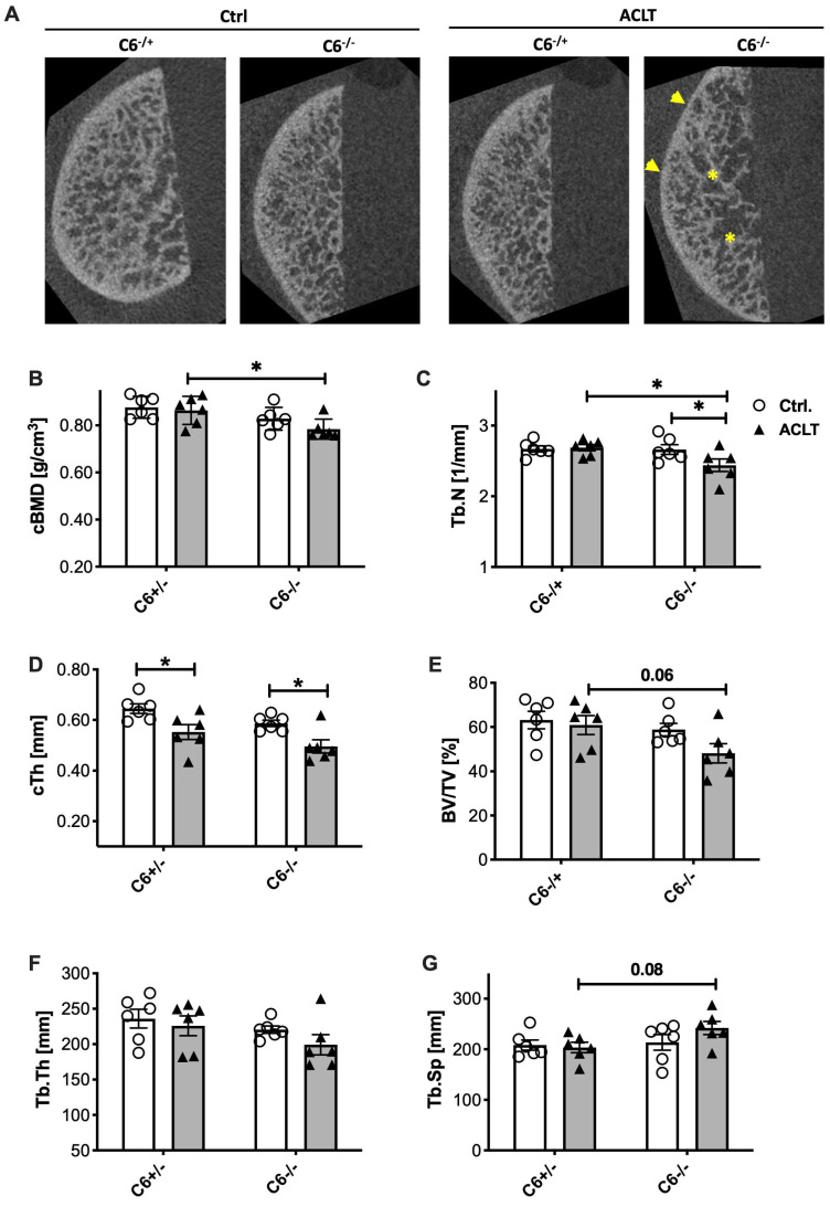
The terminal complement complex (TCC) has been described as a potential driver in the pathogenesis of posttraumatic osteoarthritis (PTOA). However, sublytic TCC deposition might also play a crucial role in bone development and regeneration. Therefore, we elucidated the effects of TCC on joint-related tissues using a rabbit PTOA model. In brief, a C6-deficient rabbit breed was characterized on genetic, protein, and functional levels. Anterior cruciate ligament transection (ACLT) was performed in C6-deficient (C6-/-) and C6-sufficient (C6+/-) rabbits. After eight weeks, the progression of PTOA was determined histologically. Moreover, the structure of the subchondral bone was evaluated by µCT analysis. C6 deficiency could be attributed to a homozygous 3.6 kb deletion within the C6 gene and subsequent loss of the C5b binding site. Serum from C6-/- animals revealed no hemolytic activity. After ACLT surgery, joints of C6-/- rabbits exhibited significantly lower OA scores, including reduced cartilage damage, hypocellularity, cluster formation, and osteophyte number, as well as lower chondrocyte apoptosis rates and synovial prostaglandin E2 levels. Moreover, ACLT surgery significantly decreased the trabecular number in the subchondral bone of C6-/- rabbits. Overall, the absence of TCC protected from injury-induced OA progression but had minor effects on the micro-structure of the subchondral bone.
Keywords: ACLT; C6; PTOA; TCC; complement activation; osteoarthritis; terminal complement complex.
Conflict of interest statement
The authors declare no conflict of interest.
Figures
Similar articles
-
Terminal complement complex deposition on chondrocytes promotes premature senescence in age- and trauma-related osteoarthritis.Front Immunol. 2025 Jan 14;15:1470907. doi: 10.3389/fimmu.2024.1470907. eCollection 2024. Front Immunol. 2025. PMID: 39877352 Free PMC article.
-
[In vitro effect of alendronate on chondrocytes and articular cartilage and subchondral bone in rabbit anterior cruciate ligament transection model].Zhongguo Xiu Fu Chong Jian Wai Ke Za Zhi. 2009 Dec;23(12):1474-81. Zhongguo Xiu Fu Chong Jian Wai Ke Za Zhi. 2009. PMID: 20073314 Chinese.
-
Inhibition of cartilage degradation and suppression of PGE2 and MMPs expression by pomegranate fruit extract in a model of posttraumatic osteoarthritis.Nutrition. 2017 Jan;33:1-13. doi: 10.1016/j.nut.2016.08.004. Epub 2016 Sep 2. Nutrition. 2017. PMID: 27908544 Free PMC article.
-
A Systematic Review of Basic Science and Animal Studies on the Use of Doxycycline to Reduce the Risk of Posttraumatic Osteoarthritis After Anterior Cruciate Ligament Rupture/Transection.Am J Sports Med. 2021 Jul;49(8):2255-2261. doi: 10.1177/0363546520965971. Epub 2020 Nov 20. Am J Sports Med. 2021. PMID: 33216621
-
The role of subchondral bone damage in post-traumatic osteoarthritis.Ann N Y Acad Sci. 2016 Nov;1383(1):58-66. doi: 10.1111/nyas.13261. Epub 2016 Sep 26. Ann N Y Acad Sci. 2016. PMID: 27671712 Review.
Cited by
-
Emerging concepts and challenges in the development of disease-modifying osteoarthritis drugs - a more refined perspective.Arch Pharm Res. 2025 Jun;48(6):467-494. doi: 10.1007/s12272-025-01551-3. Epub 2025 Jun 28. Arch Pharm Res. 2025. PMID: 40580372 Free PMC article. Review.
-
Analysis of Intervertebral Disc Degeneration Induced by Endplate Drilling or Needle Puncture in Complement C6-Sufficient and C6-Deficient Rabbits.Biomedicines. 2024 Jul 30;12(8):1692. doi: 10.3390/biomedicines12081692. Biomedicines. 2024. PMID: 39200157 Free PMC article.
-
Terminal complement complex deposition on chondrocytes promotes premature senescence in age- and trauma-related osteoarthritis.Front Immunol. 2025 Jan 14;15:1470907. doi: 10.3389/fimmu.2024.1470907. eCollection 2024. Front Immunol. 2025. PMID: 39877352 Free PMC article.
References
-
- Carpanini S.M., Torvell M., Bevan R.J., Byrne R.A.J., Daskoulidou N., Saito T., Saido T.C., Taylor P.R., Hughes T.R., Zelek W.M., et al. Terminal complement pathway activation drives synaptic loss in Alzheimer's disease models. Acta Neuropathol. Com. 2022;10:99. doi: 10.1186/s40478-022-01404-w. - DOI - PMC - PubMed
Publication types
MeSH terms
Substances
LinkOut - more resources
Full Text Sources
Medical
