. 2023 Feb 3;13(2):287.
doi: 10.3390/biom13020287.

Copper Binding and Redox Activity of α-Synuclein in Membrane-Like Environment

Affiliations

Copper Binding and Redox Activity of α-Synuclein in Membrane-Like Environment

Chiara Bacchella et al. Biomolecules. .

Abstract

α-Synuclein (αSyn) constitutes the main protein component of Lewy bodies, which are the pathologic hallmark in Parkinson's disease. αSyn is unstructured in solution but the interaction of αSyn with lipid membrane modulates its conformation by inducing an α-helical structure of the N-terminal region. In addition, the interaction with metal ions can trigger αSyn conformation upon binding and/or through the metal-promoted generation of reactive oxygen species which lead to a cascade of structural alterations. For these reasons, the ternary interaction between αSyn, copper, and membranes needs to be elucidated in detail. Here, we investigated the structural properties of copper-αSyn binding through NMR, EPR, and XAS analyses, with particular emphasis on copper(I) coordination since the reduced state is particularly relevant for oxygen activation chemistry. The analysis was performed in different membrane model systems, such as micellar sodium dodecyl sulfate (SDS) and unilamellar vesicles, comparing the binding of full-length αSyn and N-terminal peptide fragments. The presence of membrane-like environments induced the formation of a copper:αSyn = 1:2 complex where Cu+ was bound to the Met1 and Met5 residues of two helical peptide chains. In this coordination, Cu+ is stabilized and is unreactive in the presence of O2 in catechol substrate oxidation.

Keywords: copper(I); copper(II); membrane environment; redox activity; synuclein; α-helix.


Conflict of interest statement

The authors declare no conflict of interest.

Figures

Figure 1
Figure 1
Overlaid 1H-15N HSQC spectra of micelle-bound αSyn1–140 (15N uniformly enriched) in the presence of increasing amounts of metal ion: (A) 0 eq (black), 0.3 Ag+ equiv. (light blue), 0.6 Ag+ equiv. (magenta); (B) 0.6 Ag+ equiv. (black), 0.8 Ag+ equiv. (light blue), 1 Ag+ equiv. (orange); (C) 0 eq (black), 0.2 Cu2+ equiv. (light blue), 0.4 Cu2+ equiv. (red), 0.6 Cu2+ equiv. (green). αSyn1–140 240 μM, SDS-d25 50 mM, phosphate buffer 20 mM pH = 7.4, T = 298 K.
Figure 2
Figure 2
Cu K-edge XANES spectra of the membrane-bound αSyn1–140 and αSyn1–15 Cu+ complexes. αSyn1–140 500 μM, αSyn1–15 500 μM, Cu2+ 250 μM, SDS 50 mM, 125:1 lipid:protein ratio, phosphate buffer 20 mM pH = 7.4.
Figure 3
Figure 3
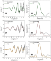
Experimental and simulated k3 weighted EXAFS spectra of αSyn1–15 SDS-Cu+ (1A), αSyn SDS-Cu+ (2A), and αSyn UVs-Cu+ (3A), and Fourier transform of αSyn1–15 SDS-Cu+ (1B), αSyn SDS-Cu+ (2B), and αSyn UVs-Cu+ (3B) spectra. αSyn1–140 500 μM, αSyn1–15 500 μM, Cu2+ 250 μM, SDS 50 mM, 125:1 lipid:protein ratio, phosphate buffer 20 mM pH = 7.4.
Figure 4
Figure 4
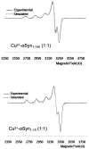
EPR spectra of micelle-bound synuclein Cu2+ complexes: αSyn1–140 (upper panel) and αSyn1–15 (lower panel). αSyn1–140 500 μM, αSyn1–15 500 μM, Cu2+ 500 μM, SDS 50 mM, phosphate buffer 20 mM pH = 7.4.
Figure 5
Figure 5
Kinetic profiles of 4-MC (3 mM) oxidation over time in 50 mM HEPES buffer at pH 7.4 and 20 °C containing SDS (20 mM) and in the presence of Cu+ (25 μM) alone (orange trace) or Cu+ (25 μM) and αSyn (50 μM) (grey trace). Copper(I) was generated in situ by anaerobic reaction of copper(II) nitrate (25 μM) and ascorbate (50 μM) prior to exposure of the solution to air.
Figure 6
Figure 6
Schematic representations of αSyn copper(II)-binding sites in aqueous and membrane environments.
Figure 7
Figure 7
Schematic representations of αSyn copper(I)-binding sites in aqueous and membrane environments.

References

    1. Wakabayashi K., Tanji K., Mori F., Takahashi H. The Lewy body in Parkinson’s disease: Molecules implicated in the formation and degradation of alpha-synuclein aggregates. Neuropathology. 2007;27:494–506. doi: 10.1111/j.1440-1789.2007.00803.x. - DOI - PubMed
    1. Runwal G., Edwards R.H. The Membrane Interactions of Synuclein: Physiology and Pathology. Annu. Rev. Pathol. 2021;16:465–485. doi: 10.1146/annurev-pathol-031920-092547. - DOI - PubMed
    1. Burré J., Sharma M., Tsetsenis T., Buchman V., Etherton M.R., Südhof T.C. Alpha-synuclein promotes SNARE-complex assembly in vivo and in vitro. Science. 2010;329:1663–1667. doi: 10.1126/science.1195227. - DOI - PMC - PubMed
    1. Bartels T., Ahlstrom L.S., Leftin A., Kamp F., Haass C., Brown M.F., Beyer K. The N-terminus of the intrinsically disordered protein α-synuclein triggers membrane binding and helix folding. Biophys. J. 2010;99:2116–2124. doi: 10.1016/j.bpj.2010.06.035. - DOI - PMC - PubMed
    1. Hashimoto M., Takenouchi T., Mallory M., Masliah E., Takeda A. The role of NAC in amyloidogenesis in Alzheimer’s disease. Am. J. Pathol. 2000;156:734–736. doi: 10.1016/S0002-9440(10)64777-3. - DOI - PMC - PubMed

Publication types