. 2023 Feb 11;13(2):352.
doi: 10.3390/biom13020352.

Proton Migration on Top of Charged Membranes

Affiliations

Proton Migration on Top of Charged Membranes

Ewald Weichselbaum et al. Biomolecules. .

Abstract

Proton relay between interfacial water molecules allows rapid two-dimensional diffusion. An energy barrier, ΔGr‡, opposes proton-surface-to-bulk release. The ΔGr‡-regulating mechanism thus far has remained unknown. Here, we explored the effect interfacial charges have on ΔGr‡'s enthalpic and entropic constituents, ΔGH‡ and ΔGS‡, respectively. A light flash illuminating a micrometer-sized membrane patch of a free-standing planar lipid bilayer released protons from an adsorbed hydrophobic caged compound. A lipid-anchored pH-sensitive dye reported protons' arrival at a distant membrane patch. Introducing net-negative charges to the bilayer doubled ΔGH‡, while positive net charges decreased ΔGH‡. The accompanying variations in ΔGS‡ compensated for the ΔGH‡ modifications so that ΔGr‡ was nearly constant. The increase in the entropic component of the barrier is most likely due to the lower number and strength of hydrogen bonds known to be formed by positively charged residues as compared to negatively charged moieties. The resulting high ΔGr‡ ensured interfacial proton diffusion for all measured membranes. The observation indicates that the variation in membrane surface charge alone is a poor regulator of proton traffic along the membrane surface.

Keywords: bioenergetics; caged proton; fluorimetry; interfacial proton diffusion; local proton circuit; proton transport.


Conflict of interest statement

The authors declare no conflict of interest.

Figures

Figure 1
Figure 1
Representative records of interfacial proton migration. (A) Experimental scheme. Exposure of a 10 × 10 µm² area to a UV flash released protons from a caged compound that was adsorbed to the surface of a free-standing planar bilayer. The fluorescence of a membrane-anchored pH sensor in a distant 10 × 10 µm²-sized membrane patch changed upon proton arrival. (B) The changes in fluorescence intensity measured at a distance of 80 µm with negatively charged bilayers (1,2-dioleoyl-sn-glycero-3-phospho-(1′-rac-glycerol), DOPG) and uncharged bilayers (1,2-dioleoyl-sn-glycero-3-phosphocholine, DOPC) differ from each other. (C) Structural formulas of the three lipids: DOPC, DOPG, and 1,2-dioleoyl-3-trimethylammonium-propane (DOTAP). (D) The changes in fluorescence intensity at a distance of 50 μm are smaller for positively (DOTAP/DOPC mixture) charged membranes than for DOPC membranes. The four colored traces are data from representative individual release events. The buffer (pH = 9) contained 10 mM KCl.
Figure 2
Figure 2
Kinetics of proton concentration changes adjacent to the DOPG membrane surface measured at different distances x from the release spot. The temperature was equal to 19 °C. The representative colored traces represent an average of 20 individual release events. The global fits of the non-equilibrium model (Equation (8)) to all traces measured for the four distances at 19 °C (also to those traces not shown here) are depicted as solid black lines. Data amplitude was normalized to 0.5, and the individual curves were shifted vertically for representational reasons.
Figure 3
Figure 3
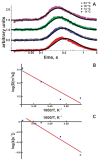
(A) Kinetics of the proton concentration adjacent to the DOPG membrane surface at 100 μm from the release spot for different temperatures. The colored traces are data from representative individual release events. The global fits of the non-equilibrium model (Equation (8)) to all traces at a given temperature (compare Figure 2) are depicted as solid black lines. Data amplitude was normalized to 0.5, and the individual curves were shifted vertically for better visibility. (B) Temperature dependence of the lateral diffusion coefficient D (in units μm2/s). The slope of the linear fit (red line) corresponds to ΔHD = (4.5 ± 1.2) kBT. We found the pre-exponential factor Ak = 6 × 10⁵ s–1. (C) Temperature dependence of the proton surface to bulk release constant, koff (in units of s−1). The slope of the linear fit (red line) corresponds to ΔHk = (13 ± 3) kBT, the intercept to AD = 1.3 × 107 μm/s2.
Figure 4
Figure 4
(A) Representative time traces of normalized proton concentration changes adjacent to the DOTAP/DOPC membrane surface at the indicated temperatures (colored traces). The distance from the release spot was equal to 50 μm. The solid black lines represent global fits of the non-equilibrium model (Equation (8)) to all traces at a given temperature. The individual curves were shifted vertically to avoid overlap. (B) Temperature dependence of the lateral diffusion coefficient (D, in units μm2/s). The slope of the temperature dependence corresponds to ΔHD = 6.6 ± 0.9 kBT. (C) The global fit of Equation (8) to the time traces (compare panel A) worked with koff set to (1.2 ± 1.2) s−1 for all measured temperatures, indicating ΔHk = 0 ± 3 kBT.

References

    1. Prins R. Hydrogen Spillover. Facts and Fiction. Chem. Rev. 2012;112:2714–2738. doi: 10.1021/cr200346z. - DOI - PubMed
    1. Kreuer K.D., Paddison S.J., Spohr E., Schuster M. Transport in proton conductors for fuel-cell applications: Simulations, elementary reactions, and phenomenology. Chem. Rev. 2004;104:4637–4678. doi: 10.1021/cr020715f. - DOI - PubMed
    1. Heberle J., Riesle J., Thiedemann G., Oesterhelt D., Dencher N.A. Proton migration along the membrane surface and retarded surface to bulk transfer. Nature. 1994;370:379–382. doi: 10.1038/370379a0. - DOI - PubMed
    1. Alexiev U., Mollaaghababa R., Scherrer P., Khorana H.G., Heyn M.P. Rapid long-range proton diffusion along the surface of the purple membrane and delayed proton transfer into the bulk. Proc. Natl. Acad. Sci. USA. 1995;92:372–376. doi: 10.1073/pnas.92.2.372. - DOI - PMC - PubMed
    1. Serowy S., Saparov S.M., Antonenko Y.N., Kozlovsky W., Hagen V., Pohl P. Structural proton diffusion along lipid bilayers. Biophys. J. 2003;84:1031–1037. doi: 10.1016/S0006-3495(03)74919-4. - DOI - PMC - PubMed

Publication types

LinkOut - more resources