Review
. 2023 Jan 26;11(2):350.
doi: 10.3390/biomedicines11020350.

Cardiac Resynchronization Therapy and Hypertrophic Cardiomyopathy: A Comprehensive Review

Affiliations
Review

Cardiac Resynchronization Therapy and Hypertrophic Cardiomyopathy: A Comprehensive Review

Andrei Dan Radu et al. Biomedicines. .

Abstract

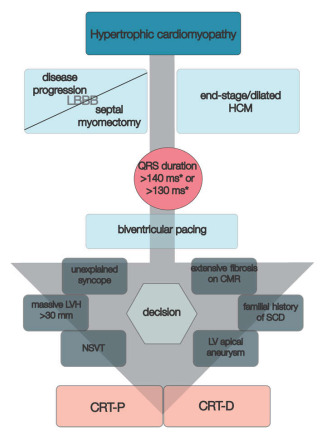
Hypertrophic cardiomyopathy (HCM) is an inherited primary myocardial disease characterized by asymmetrical/symmetrical left ventricle (LV) hypertrophy, with or without LV outflow tract (LVOT) dynamic obstruction, and poor prognosis. Cardiac resynchronization therapy (CRT) has emerged as a minimally invasive tool for patients with heart failure (HF) with decreased LV ejection fraction (LVEF) and prolonged QRS duration of over 120 ms with or without left bundle branch block (LBBB). Several HCM patients are at risk of developing LBBB because of disease progression or secondary to septal myomectomy, while others might develop HF with decreased LVEF, alleged end-stage/dilated HCM, especially those with thin myofilament mutations. Several studies have shown that patients with myectomy-induced LBBB might benefit from left bundle branch pacing or CRT to relieve symptoms, improve exercise capacity, and increase LVEF. Otherwise, patients with end-stage/dilated HCM and prolonged QRS interval could gain from CRT in terms of NYHA class improvement, LV systolic performance increase and, to some degree, LV reverse remodeling. Moreover, several electrical and imaging parameters might aid proper selection and stratification of HCM patients to benefit from CRT. Nonetheless, current available data are scarce and further studies are still required to accurately clarify the view. This review reassesses the importance of CRT in patients with HCM based on current research by contrasting and contextualizing data from various published studies.

Keywords: apical rocking; cardiac resynchronization therapy; end-stage/dilated hypertrophic cardiomyopathy; hypertrophic cardiomyopathy; left bundle branch block.


Conflict of interest statement

The authors declare no conflict of interest.

Figures

Figure 1
Figure 1
Echocardiographic findings in hypertrophic cardiomyopathy. TTE: obstructive HCM (Maron type III) in PLAX (A) and PSAX (B) views with an IVS of 23 mm and a ILW of 19 mm, with SAM of MV’s anterior leaflet in PLAX M-Mode (C) and a LVOT gradient of 127 mmHg and velocity of 5.65 m/s (D); contrast transthoracic echocardiography of a HCM with mid-cavity obstruction and apical LV aneurysm in A4C in systole (E) and diastole (F); HCM (Maron type IV) with isolated hypertrophy (18 mm) of the apical segment of LV’s lateral wall (G, H); non-obstructive HCM (Maron type II) with isolated hypertrophy of the IVS (20 mm) (I); obstructive HCM with SAM of the MV’s anterior leaflet and of its chordae tendineae with leads to severe MR (JL). TEE: obstructive HCM with SAM of MV’s anterior leaflet—A2 scallop (MO). Abbreviations: A4C, apical four-chamber view; HCM, hypertrophic cardiomyopathy; IVS, interventricular septum; LV, left ventricle; LVOT, left ventricle outflow tract; MV, mitral valve; PLAX, parasternal long-axis view; PSAX, parasternal short-axis view; SAM, systolic anterior movement; TEE, transoesophageal echocardiography; TTE, transthoracic echocardiography.
Figure 2
Figure 2
Contrast-enhanced CMR in a 50-year-old male patient with hypertrophic cardiomyopathy (from the own collection of the authors). (Upper row): b-SSFP cine images in two chambers, four chambers, short axis and three chambers, respectively. There is asymmetrical LV hypertrophy, with a maximum thickness of 20 mm at the basal anterior wall. The LV is not dilated and the LVEF is within normal limits (59%). There is systolic anterior motion of the mitral valve, resulting in acceleration of the systolic flow in the LV outflow tract. (Lower row): LGE imaging shows patchy myocardial fibrosis of the hypertrophied segments. Abbreviations: b-SSFP, balanced steady-state free precession; CMR, cardiac magnetic resonance imaging; LVEF, left ventricle ejection fraction; LGE, late gadolinium enhancement; LV, left ventricle.
Figure 3
Figure 3
Contrast-enhanced cardiovascular magnetic resonance imaging in a 32-year-old male patient with a history of hypertrophic cardiomyopathy due to a pathogenic mutation in MYBPC3 gene (c.772G>A) (from the own collection of the authors). The current findings are compatible with a phenotype of burn-out HCM. (Upper row): b-SSFP cine images in two, four and three chambers, and short axis, respectively. The LV is severely dilated (166 mL/m2) with severe systolic dysfunction (LVEF 22%). Currently, there is no LV outflow tract obstruction, although an obstructive phenotype was diagnosed 10 years before. (Lower row): LGE imaging shows extensive heterogeneous replacement myocardial fibrosis at the level of the interventricular septum, anterior and inferior LV walls, respectively. Also note the fibrosis of the papillary mitral muscles. Abbreviations: b-SSFP, balanced steady-state free precession; CMR, cardiac magnetic resonance imaging; LVEF, left ventricle ejection fraction; LGE, late gadolinium enhancement; LV, left ventricle.
Figure 4
Figure 4
Contrast-enhanced CMR in a 50-year-old female patient with a history of hypertrophic cardiomyopathy with LV apical aneurysm (from the own collection of the authors). (Upper row): b-SSFP cine images in four, three and two chambers, and short axis, respectively. The LV is dilated (110 mL/m2) with severely impaired systolic function (LVEF 25%). There is asymmetrical LV hypertrophy with a maximum wall thickness of 28 mm at the level of the interventricular septum. Note the large apical aneurysm with a maximum diameter of 40 mm and thin walls. (Lower row): LGE imaging shows transmural fibrosis of the LV apical aneurysm and a small focal intramyocardial scar at the level of the basal infero-lateral wall. The total percent of fibrosis is 16% of the LV myocardium. No thrombus is seen inside the apical aneurysm. Abbreviations: b-SSFP, balanced steady-state free precession; CMR, cardiac magnetic resonance imaging; LVEF, left ventricle ejection fraction; LGE, late gadolinium enhancement; LV, left ventricle.
Figure 5
Figure 5
Clinical vignette of LBBB and post-CRT electrical patterns and post-procedural radiological anatomy in patients with hypertrophic cardiomyopathy treated with CRT (own collection of authors). (A)—typical LBBB activation; (B)—prior AAIR pacing for sinus node disease with atypical LBBB activation; (C)—atypical LBBB with apical aneurysm in midventricular hypertrophy pattern HCM; (D)—DDDR programming with LV-only pacing in optimal fusion intervals in an HCM patient with LBBB; (E)—DDD programming with biventricular pacing in an HCM patient; (F)—post-procedural radiological aspect of CRT-D device with mid-septal position of single-coil RV lead and posterolateral position of LV bipolar lead in a non-dilated hypokinetic HCM patient; (G)—post-procedural radiological aspect of a preexisting single-chamber ICD upgraded to CRT-D with a mid-septal position of dual-coil RV lead and posterolateral position of LV tetrapolar lead in a patient with end-stage dilated phenotype of HCM with severe LV dysfunction with newly developed LBBB; (H)—post-procedural radiological CRT aspect with mid-septal position of single-coil RV lead and posterolateral position tetrapolar multipoint LV lead in a patient with prior surgical septal myectomy. Abbreviations: LBBB, left bundle branch block; CRT, cardiac resynchronization therapy; HCM, hypertrophic cardiomyopathy; RV, right ventricle; LV, left ventricle.
Figure 6
Figure 6
CRT in primary prevention of SCD in patients with hypertrophic cardiomyopathy. * > 140 ms in men and >130 ms in women. Abbreviations: CMR, cardiac magnetic resonance imaging; CRT-D, cardiac resynchronization therapy with defibrillator; CRT-P, cardiac resynchronization therapy with pacemaker; HCM, hypertrophic cardiomyopathy; LV, left ventricle; LVH, left ventricle hypertrophy; NSVT, non-sustained ventricular tachycardia; SCD, sudden cardiac death.

Similar articles

Cited by

  • A Narrative Review of Current and Investigational Therapies in Hypertrophic Cardiomyopathy.
    Ogurek I, Gill R, Tasouli-Drakou V, Joseph R, Khalid A, Houshmand N, Tak T. Ogurek I, et al. Biomedicines. 2025 May 29;13(6):1327. doi: 10.3390/biomedicines13061327. Biomedicines. 2025. PMID: 40564046 Free PMC article. Review.
  • Arrhythmias and Sudden Death: What is New in Hypertrophic Cardiomyopathy?
    Rajan D, Zörner CR, Hansen ML, Tfelt-Hansen J. Rajan D, et al. Card Fail Rev. 2025 Apr 1;11:e08. doi: 10.15420/cfr.2024.38. eCollection 2025. Card Fail Rev. 2025. PMID: 40242137 Free PMC article. Review.
  • Mexican guidelines 2024 for the diagnosis and treatment of hypertrophic cardiomyopathy.
    Llamas-Esperón GA, Berrios-Bárcenas EA, Cossío-Aranda JE, Guerra-López A, Magaña-Serrano JA, Iturralde-Torres P, Lara-Vargas JA, Meave-González A, Alexanderson-Rosas E, Ilarraza-Lomelí H, Salazar-Mendiguchía J, García-Saldivia M, Barragán-García R, Márquez-Murillo MF, Sanchez EAA, Ortiz-Ávalos M, Buendía-Hernández A, Molina-Fernández LG, Rodríguez-Diez G, Aranda-Frausto A, Gaspar-Hernández J, Eid-Lidt G, Virgen-Carrillo LR, Vásquez-Ortiz ZY, Ruiz-Esparza ME, García-Cruz E, Ávila-Vanzzini N, Vallejo-Venegas EP, Vacío-Olguín ME, Llamas-Delgado G, González-Hermosillo JA, Marmolejo-Hernández I, Sandoval-Rodríguez E, Salgado-Loza JL, Moreno-Salgado R, Chávez-Mendoza A, Martínez-Sánchez C, Ojeda-Delgado JL, Laínez-Zelaya JS, González-Romero S, Luna-Calvo MA, Gaxiola-López E, Mariona-Montero VA, Díaz CJ, Enciso-Muñoz JM, Pereira-López GI, Patrón-Chi SA, Koretzky SG, Ramírez-Moreno M. Llamas-Esperón GA, et al. Arch Cardiol Mex. 2024;94(Supl 4):1-75. doi: 10.24875/ACM.M25000098. Arch Cardiol Mex. 2024. PMID: 39928711 Free PMC article. English. No abstract available.
  • A heart failure center model helped to promote the standardized management and improve the prognosis of patients.
    Li Z, Wang Y, Hu D, Huang J, Zhang Y. Li Z, et al. Cardiovasc Diagn Ther. 2025 Feb 28;15(1):128-136. doi: 10.21037/cdt-24-437. Epub 2025 Feb 21. Cardiovasc Diagn Ther. 2025. PMID: 40115097 Free PMC article.

References

    1. Maron: B.J., Desai M.Y., Nishimura R.A., Spirito P., Rakowski H., Towbin J.A., Rowin E.J., Maron M.S., Sherrid M.V. Diagnosis and Evaluation of Hypertrophic Cardiomyopathy. J. Am. Coll. Cardiol. 2022;79:372–389. doi: 10.1016/j.jacc.2021.12.002. - DOI - PubMed
    1. Muresan I.D., Agoston-Coldea L. Phenotypes of hypertrophic cardiomyopathy: Genetics, clinics, and modular imaging. Heart Fail. Rev. 2021;26:1023–1036. doi: 10.1007/s10741-020-09931-1. - DOI - PubMed
    1. Marstrand P., Han L., Day S.M., Olivotto I., Ashley E.A., Michels M., Pereira A.C., Wittekind S.G., Helms A., Saberi S., et al. Hypertrophic Cardiomyopathy with Left Ventricular Systolic Dysfunction. Circulation. 2020;141:1371–1383. doi: 10.1161/CIRCULATIONAHA.119.044366. - DOI - PMC - PubMed
    1. Keene D., I Whinnett Z. Advances in cardiac resynchronisation therapy: Review of indications and delivery options. Heart. 2022;108:889–897. doi: 10.1136/heartjnl-2020-317171. - DOI - PubMed
    1. Glikson M., Nielsen J.C., Kronborg M.B., Michowitz Y., Auricchio A., Barbash I.M., Barrabés J.A., Boriani G., Braunschweig F., Brignole M., et al. 2021 ESC Guidelines on cardiac pacing and cardiac resynchronization therapy. Eur. Heart J. 2021;42:3427–3520. doi: 10.1093/eurheartj/ehab364. - DOI - PubMed