Review
. 2023 Jan 27;11(2):371.
doi: 10.3390/biomedicines11020371.

DNA Gyrase as a Target for Quinolones

Affiliations
Review

DNA Gyrase as a Target for Quinolones

Angela C Spencer et al. Biomedicines. .

Abstract

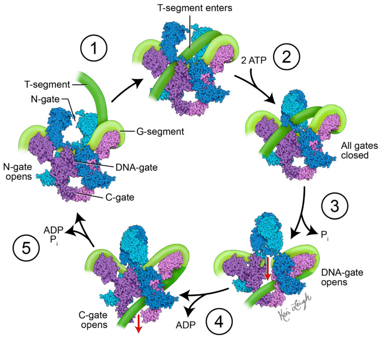
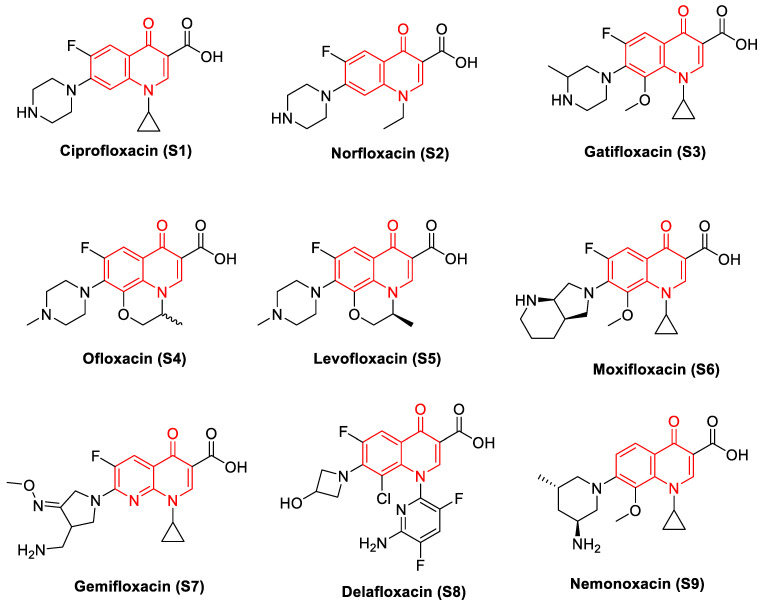
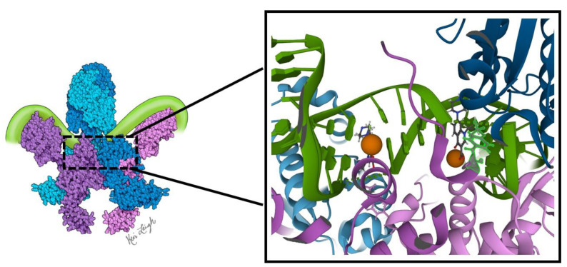
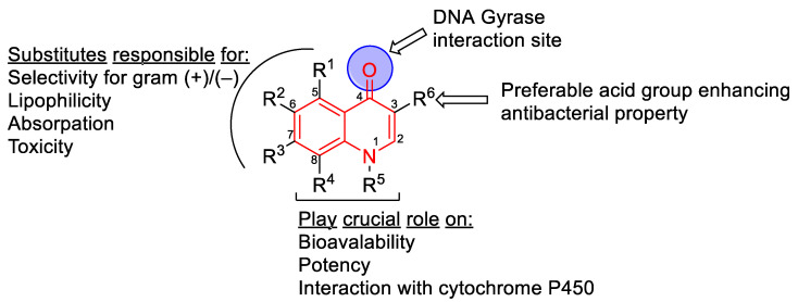
Bacterial DNA gyrase is a type II topoisomerase that can introduce negative supercoils to DNA substrates and is a clinically-relevant target for the development of new antibacterials. DNA gyrase is one of the primary targets of quinolones, broad-spectrum antibacterial agents and are used as a first-line drug for various types of infections. However, currently used quinolones are becoming less effective due to drug resistance. Common resistance comes in the form of mutation in enzyme targets, with this type being the most clinically relevant. Additional mechanisms, conducive to quinolone resistance, are arbitrated by chromosomal mutations and/or plasmid-gene uptake that can alter quinolone cellular concentration and interaction with the target, or affect drug metabolism. Significant synthetic strategies have been employed to modify the quinolone scaffold and/or develop novel quinolones to overcome the resistance problem. This review discusses the development of quinolone antibiotics targeting DNA gyrase to overcome bacterial resistance and reduce toxicity. Moreover, structural activity relationship (SAR) data included in this review could be useful for the development of future generations of quinolone antibiotics.

Keywords: DNA gyrase; drug development; drug-resistance; molecular docking; quinolones.


Conflict of interest statement

The authors declare no conflict of interest.

Figures

Figure 1
Figure 1
Proposed mechanism of action of DNA gyrase. Initially, DNA gyrase is bound to a G-segment of DNA at the DNA gate, a region located at the interface between subunits A (purple and pink) and B (blue and teal). The G-segment of DNA (light green) is wrapped around the C-terminal domains (CTDs) of the A subunits. The mechanistic steps are as follows: (1) the T-segment of DNA (dark green) enters the gyrase via the N-gate formed by the two B subunits. (2) When two ATP molecules bind the B subunits, the N-gate closes, and the T-segment DNA is trapped in the upper cavity of the gyrase. (3) The G-segment is cleaved, opening the DNA gate and allowing the T-segment to pass through into the lower cavity. This step requires the hydrolysis of one ATP with the release of Pi. (4) The C-gate opens, and the T-segment passes through with the release of ADP. The second ATP is hydrolyzed. (5) The release of ADP and Pi, along with the closing of the C-gate and opening of the N-gate, readies DNA gyrase for another cycle of supercoiling. The diagram is based on Soczek et al. [24]. The structure of DNA gyrase was derived from PDB ID 6RKW [25]. Figure created by medical illustrator Keri Leigh Jones, MSMI, CMI.
Figure 2
Figure 2
Quinolones (the scaffold colored in red): DNA gyrase poisons used as antibacterial agents.
Figure 3
Figure 3
Binding of quinolones to DNA gyrase. The quinolone ciprofloxacin is shown bound to Mycobaterium tuberculosis DNA gyrase at the cleavage core in the dimer interface. Two quinolones (shown as ball and stick, color cpk) intercalate into the G-segment DNA (green) near the cleavage site. The noncatalytic Mg2+ ion that is coordinated by the keto acid group is shown in orange. The key active site tyrosines in the A subunits are shown as ball and stick in pink and purple. The structure shown on the right is adapted from PDB ID 5BTC. The color scheme is the same as in Figure 1.
Figure 4
Figure 4
Quinolone core structure with six possible substitution locations for modification. The roles of substituents in biological activity are indicated.

Similar articles

Cited by

References

    1. Hutchings M., Truman A., Wilkinson B. Antibiotics: Past, present and future. Curr. Opin. Microbiol. 2019;51:72–80. doi: 10.1016/j.mib.2019.10.008. - DOI - PubMed
    1. Naghavi M. Global burden of bacterial antimicrobial resistance in 2019: A systematic analysis. Lancet. 2022;399:629–655. - PMC - PubMed
    1. O’Neill J. Tackling Drug-Resistant Infections Globally: Final Report and Recommendations. Government of the United Kingdom; London, UK: 2016. Review on Antimicrobial Resistance.
    1. CDC . COVID-19: U.S. Impact on Antimicrobial Resistance, Special Report 2022. U.S. Department of Health and Human Services, CDC; Atlanta, GA, USA: 2022.
    1. Shafran N., Shafran I., Ben-Zvi H., Sofer S., Sheena L., Krause I., Shlomai A., Goldberg E., Sklan E.H. Secondary bacterial infection in COVID-19 patients is a stronger predictor for death compared to influenza patients. Sci. Rep. 2021;11:12703. doi: 10.1038/s41598-021-92220-0. - DOI - PMC - PubMed

LinkOut - more resources