Review
. 2023 Feb 6;11(2):471.
doi: 10.3390/biomedicines11020471.

Peptides That Block RAS-p21 Protein-Induced Cell Transformation

Affiliations
Review

Peptides That Block RAS-p21 Protein-Induced Cell Transformation

Matthew R Pincus et al. Biomedicines. .

Abstract

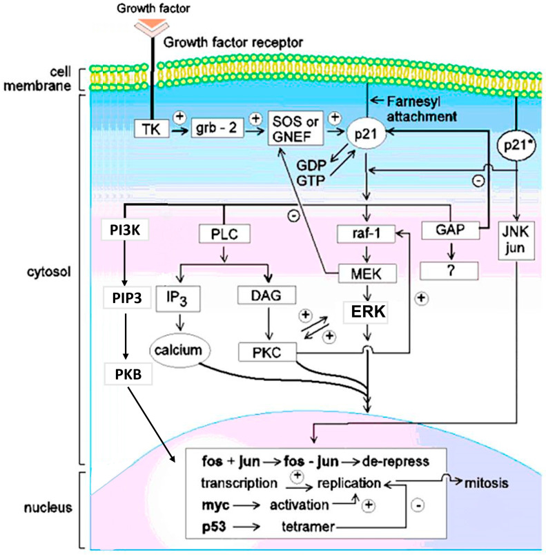
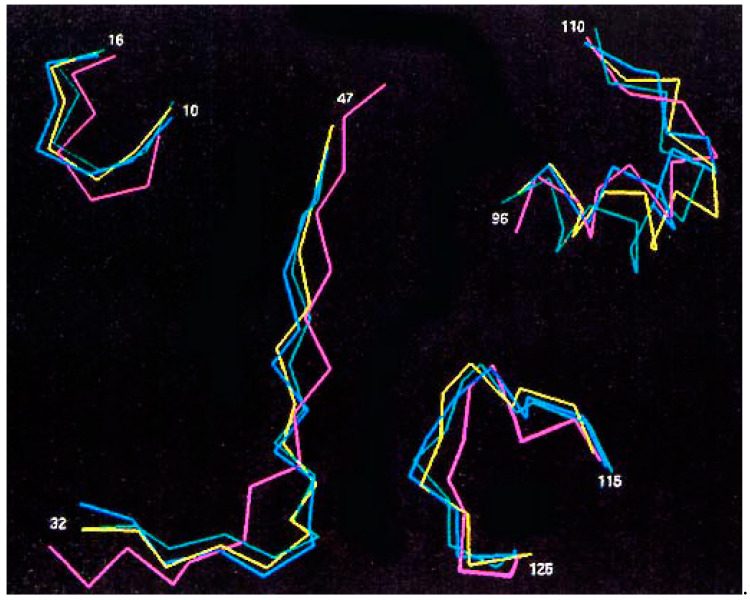
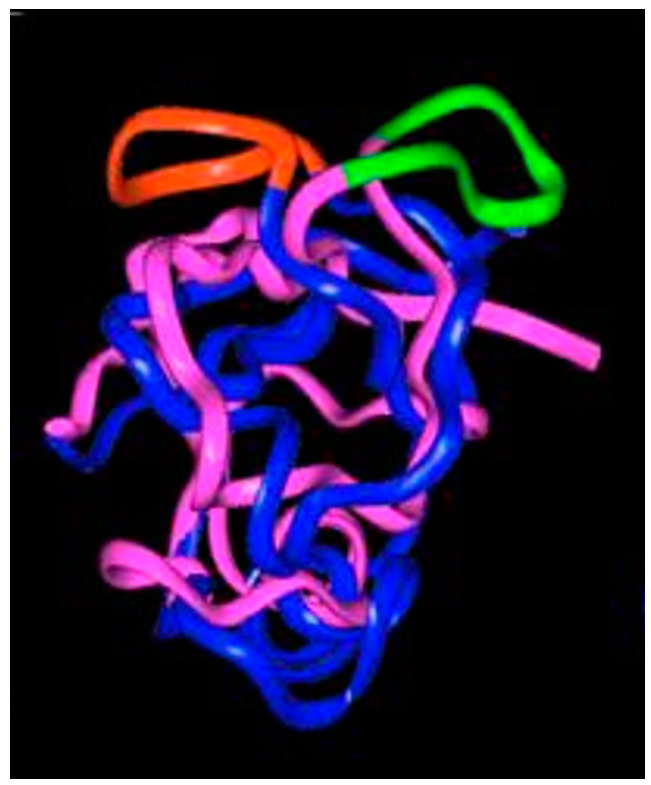
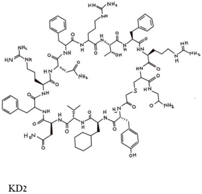
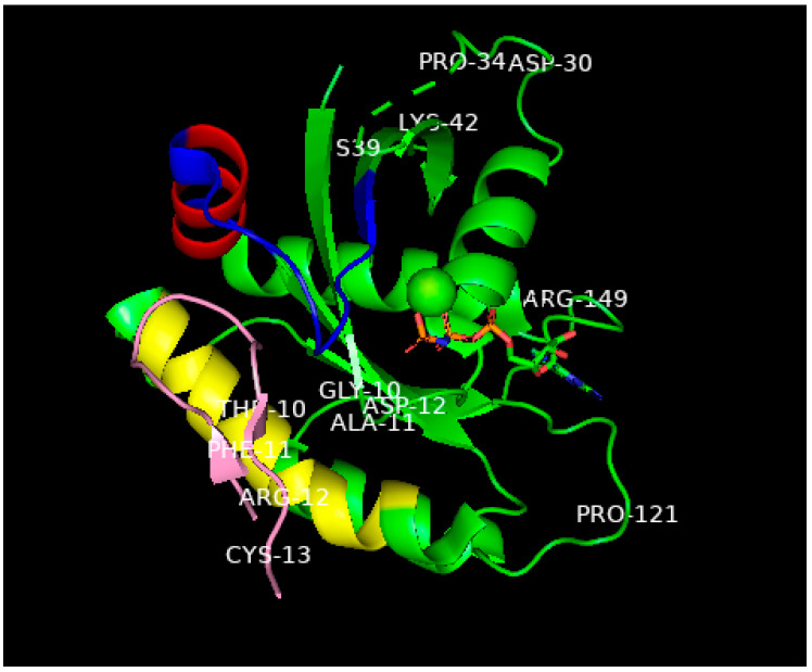
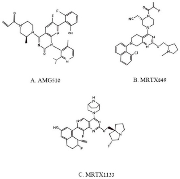
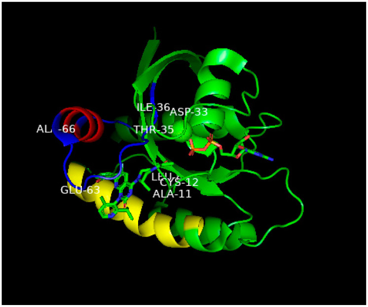
This is a review of approaches to the design of peptides and small molecules that selectively block the oncogenic RAS-p21 protein in ras-induced cancers. Single amino acid substitutions in this protein, at critical positions such as at Gly 12 and Gln 61, cause the protein to become oncogenic. These mutant proteins cause over 90 percent of pancreatic cancers, 40-50 percent of colon cancers and about one third of non-small cell cancers of the lung (NSCCL). RAS-p21 is a G-protein that becomes activated when it exchanges GDP for GTP. Several promising approaches have been developed that target mutant (oncogenic) RAS-p21 proteins in these different cancers. These approaches comprise: molecular simulations of mutant and wild-type proteins to identify effector domains, for which peptides can be made that selectively inhibit the oncogenic protein that include PNC-1 (ras residues 115-126), PNC-2 (ras residues 96-110) and PNC7 (ras residues 35-47); the use of contiguous RAS-p21 peptide sequences that can block ras signaling; cyclic peptides from large peptide libraries and small molecule libraries that can be identified in high throughput assays that can selectively stabilize inactive forms of RAS-p21; informatic approaches to discover peptides and small molecules that dock to specific domains of RAS-p21 that can block mitogenic signal transduction by oncogenic RAS-p21; and the use of cell-penetrating peptides (CPPs) that are attached to the variable domains of the anti-RAS-p21 inactivating monoclonal antibody, Y13 259, that selectively enters oncogenic RAS-p21-containing cancer cells, causing these cells to undergo apoptosis. Several new anti-oncogenic RAS-p21 agents, i.e., Amgen's AMG510 and Mirati Therapeutics' MRTX849, polycyclic aromatic compounds, have recently been FDA-approved and are already being used clinically to treat RAS-p21-induced NSCCL and colorectal carcinomas. These new drugs target the inactive form of RAS-p21 bound to GDP with G12C substitution at the critical Gly 12 residue by binding to a groove bordered by specific domains in this mutant protein into which these compounds insert, resulting in the stabilization of the inactive GDP-bound form of RAS-p21. Other peptides and small molecules have been discovered that block the G12D-RAS-p21 oncogenic protein. These agents can treat specific mutant protein-induced cancers and are excellent examples of personalized medicine. However, many oncogenic RAS-p21-induced tumors are caused by other mutations at positions 12, 13 and 61, requiring other, more general anti-oncogenic agents that are being provided using alternate methods.

Keywords: RAS-p21 protein; amino acid substitutions; blockade of oncogenic protein; cell transformation; mutant protein; oncogenic forms; peptides; small molecules.


Conflict of interest statement

None of the authors of this paper have any real or perceived conflicts of interest concerning any aspects of the work presented in this paper.

Figures

Figure 1
Figure 1
Pincus et al. Wild-type (RAS-p21) and Oncogenic (RAS-p21*) Signal Transduction Pathways. A growth factor such as EGF or insulin binds to its receptor (upper left)—for example, EGFR—extracellularly. The receptor contains three domains: the extracellular growth factor-binding domain, the trans-membrane domain and the intracytosolic domain. The latter contains a tyrosine kinase (TK in the figure) which becomes activated when the receptors dimerize upon binding to the growth factor. This event enables the intracytosolic domain to bind to the adapter grb-2 protein that concurrently binds to and activates a guanine nucleotide exchange factor (GNEF), SOS, a protein that in turn binds to RAS-p21, causing it to exchange GDP for GTP. In the GTP-bound state, RAS-p21 becomes activated. In its activated state, it induces mitogenic signal transduction which occurs only if RAS-p21 is bound to the inner surface of the cell membrane through a covalently attached farnesyl moiety in thioether linkage to Cys 186 catalyzed by the enzyme, farnesyl transferase. If RAS-p21 is not membrane-bound, it acts as a competitive inhibitor of membrane-bound ras. An important target of activated membrane-bound RAS-p21 in mitogenic signaling is the serine-theonine kinase, raf, a 74 kDa Ser/Thr kinase protein that, in turn, directly binds to and activates another serine-threonine kinase, MEK in a so-called kinase cascade. The latter protein activates ERK. This all-important protein shuttles between the cytosol, where it is involved in cytoskeletal rearrangements (the name MAPK stands for mitogen activated or microtubule-activating protein kinase) and the nucleus in which it activates the critically important nuclear transcription factor, fos, that itself forms a heterodimeric complex, called AP1, with the critically important nuclear transcription factor, jun, that is activated by another kinase, jun-N-terminal kinase (JNK), that is normally activated on a separate pathway called the stress-activated protein (SAP) pathway. The AP1 complex induces the transcription of many pro-mitogenic proteins including cyclins and nuclear skeletal proteins called nuclear matrix proteins ( or NMP’s); other nuclear proteins, such as myc, also transcriptionally active, are also often activated in this process. Anti-oncogene proteins, such as p53, also become activated. This protein blocks the transcription of pro-mitotic proteins (minus sign on p53 arrow) and induces apoptosis in transformed cells. On the right of the figure, oncogenic RAS-p21 (RAS-p21*) is shown interacting with and activating JNK, which seems to be a unique aspect to oncogenic RAS-p21. Work in oocytes (see text) suggests that wild-type RAS-p21 may also activate the dual specificity kinase TOPK (not shown in the figure), which is known to bind to RAF and is in the MEK superfamily. Apparently, oncogenic RAS-p21 does not utilize this pathway. The activated state of RAS-p21 is regulated by the protein, GAP (GTPase-Activating Protein), that enhances the native GTPase activity of RAS-p21. GAP induces the hydrolysis of GTP to GDP, resulting in the inactivation of RAS-p21. However, GAP itself may be involved in mitogenic signal transduction, hence, the question mark in the arrow below the GAP box in the figure. As shown on the left side of the figure, RAS-p21 also interacts directly with phosphoinositol-3-hydroxy kinase (PI3K), which catalyzes the synthesis of phosphatidylinositol 3-hydroxyphosphate or triphosphate (PIP3) that is involved in the activation of protein kinase B (PKB), also known as AkT, of which among many functions, induces cell proliferation. Activated RAS-p21 further induces the activation of phospholipase C (PLC) that catalyzes the formation of diacylglycerol (DAG), an activator of protein kinase C (PKC) that is especially critical to the oncogenic RAS-p21 pathway and the synthesis of inositol triphosphate (IP3) of which mobilizes the release of calcium.
Figure 2
Figure 2
Cα traces for effector domains of molecular dynamics average structures of different forms of RAS-p21. Color scheme is: pink, wild-type RAS-p21-GDP; yellow, RAS-p21-GTP; blue, G12V-RAS-p21-GTP; green, Q61L-RAS-p21-GTP. The residue numbers for each domain are shown in the figure [3].
Figure 3
Figure 3
Ribbon representation of the superimposed average molecular dynamics RAF RBD structures bound to oncogenic (Val 12-) RAS-p21 (blue and red structure) and to wild-type RAS-p21 (pink and green). The exposed loop on the top surface of the figure of the 97–110 segment is colored green in the wild-type complex and red in the oncogenic complex (adapted from [15]).
Figure 4
Figure 4
Pincus et al. Structure of the cyclic peptide, KD2, that blocks the activation of the G12D-RAS-p21-GTP complex. The Thr 10 residue whose side chain -OH group makes contact with the Asp 12 residue is in the top middle of the figure and can be identified by the -OH in this area.
Figure 5
Figure 5
Ribbon representation of the X-ray structure (ref. [29], PDB entry 6WGN) of KD2 bound to G12D-RAS-p21-GppNHp rotated to show the location of KD2 (pink) in the groove in the switch 2 domain (residues 55–76) which is colored blue except for the α2 helix involving residues 67–73, colored red. This helix is rotated by 90° relative to its position in wild-type RAS-p21. The yellow-colored segment is the α3 helix involving residues 89–103. A significant segment of the pink KD2 peptide is seen to bind in a deep groove between the α2 helix and a significant part of the switch 2 domain shown as the blue strand to the right and above the pink segment. GppNHp is seen in the middle, right of the figure with the guanine ring on the far right; the β- and ϒ-phosphates (the phosphates are colored orange) are seen to bind to the magnesium ion shown as a green sphere in the center of the figure. The oncogenic Asp 12 residue is shown in the middle of the P loop domain, some of whose other residues (Gly 10 and Ala 11) are also shown. Additionally, to be noted: The all-important switch 1 domain containing residues 32–47 that contacts the RBD of RAF is shown in the upper right of the figure. Some of the residues in this domain such as Pro 34, Ser 39 and Lys 42 are shown to help identify this domain. Between Pro 34 and Ser 39, there is a series of green dashes denoting the absence of location of the atoms in the intervening segment, this is due to the disordered switch 1 domain in state 1 (inactive) in GTP-bound RAS-p21. See text for explanation.
Figure 6
Figure 6
Pincus et al. Chemical structures of three poly aromatic ring molecules, AMG500 (A), MRTX849 (B) and MRTX 1139 (C) that insert into a groove formed by the switch 2 domain, its α2 helix and its α3 helix. The first two compounds, (A,B), bind uniquely to G12C-K-RAS-p21-GDP, the third, (C), binds to G12D-K-RAS-p21-GDP.
Figure 7
Figure 7
Ribbon representation of the X-ray structure of AMG510 bound to G12C-RAS-p21-GDP from Ref. [36]. Coordinates were obtained from the Protein Data Base (PDB) identity number 7RPZ and rotated to give a clear view of the binding pocket occupied by AMG510. The color scheme for critical domains is the same as for Figure 5. G12C-RAS-p21 uniquely has a groove formed by the switch 2 domain, consisting of residues 55–76 (colored blue except for its α2 helix that is colored red) and its α3 helix (residues 89–103, colored yellow). The orientation of this complex is similar to that of the KD2-G12D-RAS-p21-GppNHp complex shown in Figure 5. As in Figure 5, the nucleotide (here, GDP) is oriented horizontally such that the guanine ring is on the far right in the middle of the figure, and a β-phosphate oxygen binds to the magnesium ion (green sphere in the middle of the figure). The AMG510 molecule can be seen in the lower left of the protein inserted into a groove between the yellow α3 helix and the blue switch 2 loop region above it in the figure. Just above Leu 80 (“Leu” in the middle of the figure), there is a red projection that is the carbonyl oxygen of the acryl moiety of the drug that is positioned to react with the-SH group of Cys 12 labeled in the figure. In contrast to Figure 5, the switch 1 domain is seen to be well-defined and can be identified by the labeled residue positions Asp 33, Thr 35 and Leu 36. Unlike the positioning of switch 1 residues on the surface of the protein in Figure 5, the switch 1 residues of G12C-RAS-p21-GDP are not as exposed.

Similar articles

Cited by

References

    1. Pincus M.R., Bluth M., Brandt-Rauf P.W., Bowne W., LaDoulis C. Oncoproteins and Early Tumor Detection, Chapter 77. In: McPherson R.A., Pincus M.R., editors. Henry’s Clinical Diagnosis and Management by Laboratory Methods. 24th ed. Elsevier; New York, NY, USA: Philadelphia, PA, USA: 2021. pp. 1525–1541.
    1. Barbacid M. ras Genes. Ann. Rev. Biochem. 1987;56:779–827. doi: 10.1146/annurev.bi.56.070187.004023. - DOI - PubMed
    1. Stacey D.W., Kung H.-F. Transformation of NIH 3T3 Cells by Microinjection of Ha-ras p21 Protein. Nature. 1984;310:508–511. doi: 10.1038/310508a0. - DOI - PubMed
    1. Birchmeier C., Broeck D., Wigler M. RAS Proteins Can Induce Meiosis in Xenopus Oocytes. Cell. 1985;43:615–621. doi: 10.1016/0092-8674(85)90233-8. - DOI - PubMed
    1. Deshpande A.K., Kung H.-F. Insulin Induction of Xenopus laevis Oocyte Maturation Is Inhibited by Monoclonal Antibody Against p21 ras Proteins. Mol. Cell Biol. 1987;7:1285–1288. - PMC - PubMed

LinkOut - more resources