Review
. 2023 Feb 7;12(4):530.
doi: 10.3390/cells12040530.

PD-1/PD-L1 and DNA Damage Response in Cancer

Affiliations
Review

PD-1/PD-L1 and DNA Damage Response in Cancer

Mateusz Kciuk et al. Cells. .

Abstract

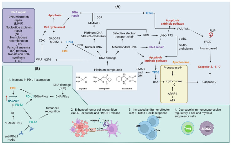
The application of immunotherapy for cancer treatment is rapidly becoming more widespread. Immunotherapeutic agents are frequently combined with various types of treatments to obtain a more durable antitumor clinical response in patients who have developed resistance to monotherapy. Chemotherapeutic drugs that induce DNA damage and trigger DNA damage response (DDR) frequently induce an increase in the expression of the programmed death ligand-1 (PD-L1) that can be employed by cancer cells to avoid immune surveillance. PD-L1 exposed on cancer cells can in turn be targeted to re-establish the immune-reactive tumor microenvironment, which ultimately increases the tumor's susceptibility to combined therapies. Here we review the recent advances in how the DDR regulates PD-L1 expression and point out the effect of etoposide, irinotecan, and platinum compounds on the anti-tumor immune response.

Keywords: DNA damage response; cytotoxic drugs; immunotherapy; programmed death ligand-1 (PD-L1).


Conflict of interest statement

The authors declare no conflict of interest.

Figures

Figure 1
Figure 1
The interaction between tumor cells and immune cells. Tumor cells are detected by adaptive (comprising of B and T cells) and innate immune system (mainly natural killer (NK) cells). Effector CD4+ and cytotoxic CD8+ T cells are capable of recognizing peptide antigens presented on MHC class II or MHC class I, respectively. (A) Immunogenic cell death (ICD) is associated with the release of damage-associated molecular patterns (DAMPs) from dying cells. Such molecular patterns, whether expressed on the surface of cells or released outside of the cell, can promote tumor antigen presentation and boost adaptive immunity. Calreticulin (CRT) expressed on cancer cells interacts with the CD91 receptor on dendritic cells (DCs) and high-mobility group protein 1 (HMGB1)—a ligand for the toll-like receptor (TLR-4) receptor on DCs is released by cancer cells. Additionally, ATP released from cancer cells interacts with the P2X purinoceptor 7 (P2RX7) on DSc. (B) DCs are required for cytotoxic CD8+ T cell priming. In this process, DCs uptake antigens from tumor cells undergoing apoptosis and with the support of CD4+ helper cells present them to CD8+ T cells. Moreover, DCs can secrete tumor necrosis factor α (TNF-α,) and interleukins (IL-6, IL-8, and IL-12) that help to trigger the anticancer immune response. (C) Activated CD8+ T cells can kill cancer cells through the release of granzyme A/B (GZMA/B), perforins (PFN), or interferon gamma (INFγ) and TNFα that (D) induce cell death through the activation of the extrinsic apoptosis pathway. The extrinsic pathway is triggered with the binding of certain ligands to the TNFα receptor super family. This leads to the oligomerization of the receptor and activation of procaspase-8 through the recruitment of adaptor proteins and the formation of a death-inducing signaling complex (DISC). (E) Cancer cells reshape the tumor immune microenvironment into an immunosuppressive surrounding that is rich in regulatory T cells (Tregs), myeloid-derived suppressor cells (MDSCs), group 2 innate lymphoid cells (ILC2s), and tumor-associated macrophages (M2) that release immunosuppressive interleukins (IL-4, IL-10, IL-13, and IL-35) or transforming growth factor β (TGF-β) and up-regulate receptors (such as programmed cell death 1 ligand 1 (PD-L1)) that “hide” tumor cells from the immune recognition (not shown here). This topic has been extensively reviewed in [15,16,17] and will not be discussed in detail here. Casp—caspase; FADD—FAS-associated death domain protein; TCR—T-cell receptor. Created with BioRender.com accessed on 1 February 2023.
Figure 2
Figure 2
Effect of irinotecan and etoposide on the DNA damage (DDR) and immune response. Cancer cell (A) Irinotecan inhibits DNA topoisomerase I which leads to the formation of single-strand breaks (SSBs) and double-strand breaks (DSBs). Single-stranded DNA (ssDNA) in the replication fork that arises due to helicase activity is coated with replication protein A (RPA) protein and a mediator of DNA damage checkpoint protein 1 (MDC-1). MDC-1 serves as a platform protein for topoisomerase 2-binding protein 1 (TOPBP1), ATR-interacting protein (ATRIP), and serine/threonine-protein kinase (ATR). In contrast, DSBs that arise through the conversion of SSBs and activity of topoisomerase II inhibitors (here etoposide) are detected by the MRN complex composed of double-strand break repair protein MRE11 (MRE11), nibrin (NBS1), and DNA repair protein (RAD50). The MRN complex is involved in the recruitment and activation of ATM kinase. Activated ATR and ATM phosphorylate have multiple targets including checkpoint kinases (CHK1/2). The phosphorylation of H2AX and accumulation of MDC-1 contribute to further recruitment of ATR/ATM kinases and amplification of damage signaling. CHK1 activation leads to the phosphorylation of signal transducers and activator of transcription (STAT) proteins that control (up-regulate) the expression of interferon regulatory factor (IRF1) and the major histocompatibility complex class I molecule (MHC-I). IRF-1 works as a transcription factor for programmed death ligand-1 (PD-L1) protein. Tumor immune microenvironment (B) Topoisomerase inhibitors stimulate the anti-tumor immune response through the increased proliferation and infiltration of CD8+ cells and the production of immunostimulatory interferon gamma (INFγ). INFγ binds to and activates the interferon-gamma receptor (INFGR) that contributes to the activation of tyrosine-protein kinase JAK (JAK)/STAT signaling to confer the increased PD-L1 expression. Moreover, it also reduces the regulatory FOXP3+ T-cells and myeloid-derived suppressor cells (MDSC) that normally attenuate the anti-tumor immune response. Cells defective in double-strand break repair components (C): Inactivation of the breast cancer type 2 susceptibility protein (BRCA2) of homologous recombination (HR) pathway and X-ray repair cross-complementing protein 5/6 (KU70/80) of the non-homologous end joining (NHEJ) pathway contributes to the activation of CHK1/STAT/IRF/PD-L1 signaling. Created with BioRender.com accessed on 1 February 2023.
Figure 3
Figure 3
Cellular and immune response to platinum compounds. (A) Cellular response to platinum compounds: Platinum compounds induce DNA damage to both nuclear and mitochondrial DNA. Mitochondrial DNA damage can lead to defects in the electron transport chain that contribute to electron leakage and the formation of reactive oxygen species (ROS). ROS contribute to DNA damage in both nuclear and mitochondrial DNA in a feedback mechanism. ROS also act as molecular messengers and contribute to the activation of the Jun amino-terminal kinase (JNK) pathway that together with the P73 protein trigger apoptotic cell death. C-ABL kinase activity and mismatch repair (MMR)-proficiency are required for cell death to occur. DNA damage activates DNA damage response (DDR), as indicated by the activation of serine/threonine kinases ATM and ATR. The outcome of DDR relies heavily on cellular tumor antigen p53 (TP53) protein: (a) activation of TP53 in response to DNA damage triggers cell cycle arrest through up-regulation of cyclin-dependent kinase inhibitor 1 (WAF1/CIP1), 45kd-growth arrest, and DNA damage (GADD45) and mouse double minute 2 homolog (MDM2) or via WAF1/CIP1-mediated inhibition of cyclin-dependent kinases (CDKs); (b) subsequent DNA repair via co-operation of nucleotide excision repair (NER), MMR, homologous recombination (HR), Fanconi anemia (FA) pathway, or translesion synthesis (TLS); or (c) apoptosis induction through intrinsic apoptosis pathway followed by ROS-damage accumulation, TP53-dependent up-regulation of Bcl2-associated agonist of cell death (BAD), Diablo IAP-binding mitochondrial protein (SMAC) and serine protease HTRA2 (OMI), or through activation of FASL/FAS extrinsic apoptosis pathway. Cisplatin induces flice-like inhibitory protein (FLIP) ubiquitination and degradation, which further activate the extrinsic pathway. (B) Effect of platinum compounds on the antitumor immune response: Cyclic GMP-AMP synthase (cGAS)/stimulator of interferon genes protein (STING) pathway and extracellular signal-regulated kinase (ERK) signaling up-regulate programmed death ligand-1 (PD-L1) expression. The DNA-dependent protein kinase catalytic subunit (DNA-PKcs) up-regulated in response to double-strand breaks (DSBs) associates with PD-L1 conferring to the activation of the ERK pathway and further up-regulation of PD-L1. This, on the one hand, contributes to immune evasion, but also increase the therapeutic efficiency of anti-PD-L1 monoclonal antibodies (mAbs). Platinum compounds also enhance tumor recognition through calreticulin (CRT) exposure on cell surface and high mobility group protein 1 (HMGB1) release, induce CD4+/CD8+ T cell response, and suppress regulatory T cells (Treg) and myeloid-derived suppressor cells (MDSC). APAF-1—apoptotic protease-activating factor 1; FADD—FAS-associated death domain protein. Created with BioRender.com accessed on 1 February 2023.
Figure 4
Figure 4
Comparison of small-molecule inhibitors and monoclonal antibodies (mAbs). Based on [202]. Created with BioRender.com accessed on 1 February 2023.
Figure 5
Figure 5
The existing links between the use of chemotherapeutic drugs, DNA damage response (DDR) inhibitors, and immunotherapy with the crucial questions, advantages, and disadvantages of their use. For reference see Section 7. Created with BioRender.com accessed on 1 February 2023.

References

    1. Dillman R.O. Cancer Immunotherapy. Cancer Biother. Radiopharm. 2011;26:1–64. doi: 10.1089/cbr.2010.0902. - DOI - PubMed
    1. Davis I.D. An Overview of Cancer Immunotherapy. Immunol. Cell. Biol. 2000;78:179–195. doi: 10.1046/j.1440-1711.2000.00906.x. - DOI - PubMed
    1. Gamrekelashvili J., Krüger C., von Wasielewski R., Hoffmann M., Huster K.M., Busch D.H., Manns M.P., Korangy F., Greten T.F. Necrotic Tumor Cell Death in Vivo Impairs Tumor-Specific Immune Responses. J. Immunol. 2007;178:1573–1580. doi: 10.4049/jimmunol.178.3.1573. - DOI - PubMed
    1. McDonnell A.M., Nowak A.K., Lake R.A. Contribution of the Immune System to the Chemotherapeutic Response. Semin. Immunopathol. 2011;33:353–367. doi: 10.1007/s00281-011-0246-z. - DOI - PubMed
    1. Gardai S.J., McPhillips K.A., Frasch S.C., Janssen W.J., Starefeldt A., Murphy-Ullrich J.E., Bratton D.L., Oldenborg P.-A., Michalak M., Henson P.M. Cell-Surface Calreticulin Initiates Clearance of Viable or Apoptotic Cells through Trans-Activation of LRP on the Phagocyte. Cell. 2005;123:321–334. doi: 10.1016/j.cell.2005.08.032. - DOI - PubMed

MeSH terms