. 2023 Feb 11;12(4):582.
doi: 10.3390/cells12040582.

Transfer of Cardiac Mitochondria Improves the Therapeutic Efficacy of Mesenchymal Stem Cells in a Preclinical Model of Ischemic Heart Disease

Affiliations

Transfer of Cardiac Mitochondria Improves the Therapeutic Efficacy of Mesenchymal Stem Cells in a Preclinical Model of Ischemic Heart Disease

Marie-Luce Vignais et al. Cells. .

Abstract

Background: The use of mesenchymal stem cells (MSCs) appears to be a promising therapeutic approach for cardiac repair after myocardial infarction. However, clinical trials have revealed the need to improve their therapeutic efficacy. Recent evidence demonstrated that mitochondria undergo spontaneous transfer from damaged cells to MSCs, resulting in the activation of the cytoprotective and pro-angiogenic functions of recipient MSCs. Based on these observations, we investigated whether the preconditioning of MSCs with mitochondria could optimize their therapeutic potential for ischemic heart disease.

Methods: Human MSCs were exposed to mitochondria isolated from human fetal cardiomyocytes. After 24 h, the effects of mitochondria preconditioning on the MSCs' function were analyzed both in vitro and in vivo.

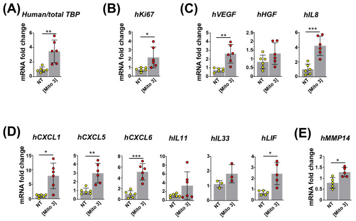
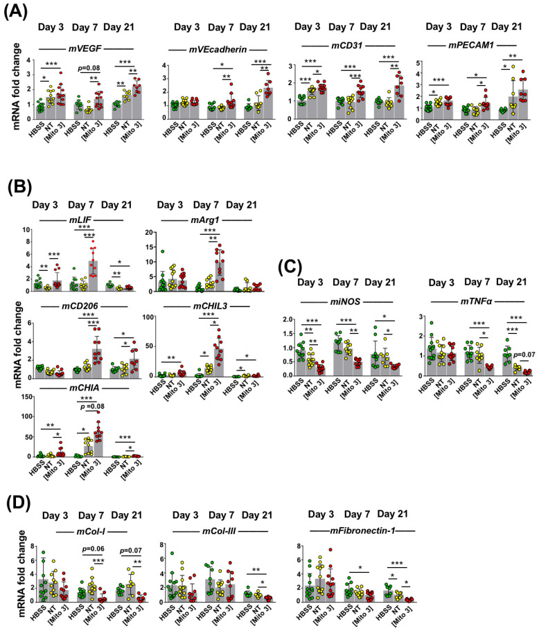
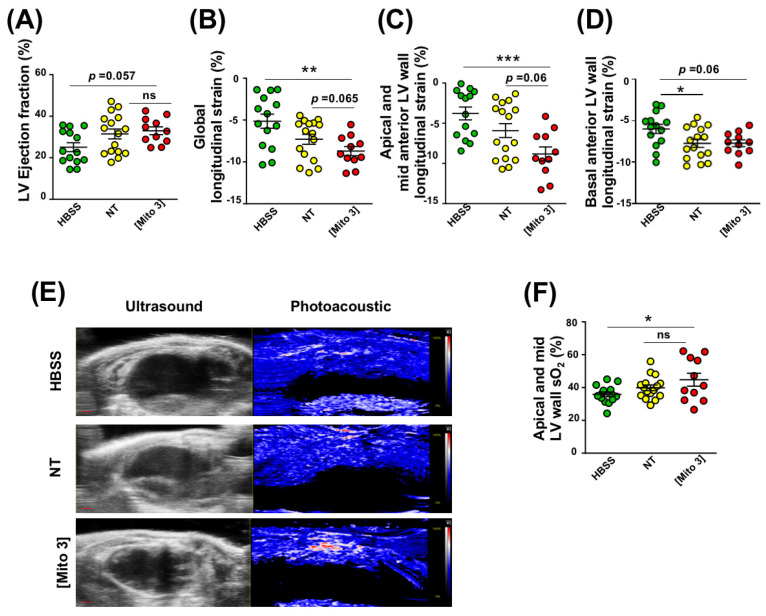
Results: We found that cardiac mitochondria-preconditioning improved the proliferation and repair properties of MSCs in vitro. Mechanistically, cardiac mitochondria mediate their stimulatory effects through the production of reactive oxygen species, which trigger their own degradation in recipient MSCs. These effects were further confirmed in vivo, as the mitochondria preconditioning of MSCs potentiated their therapeutic efficacy on cardiac function following their engraftment into infarcted mouse hearts.

Conclusions: The preconditioning of MSCs with the artificial transfer of cardiac mitochondria appears to be promising strategy to improve the efficacy of MSC-based cell therapy in ischemic heart disease.

Keywords: cell therapy; mesenchymal stem cells; metabolism; mitochondria transfer; post-ischemic heart failure.


Conflict of interest statement

The authors declare that they have no competing interests.

Figures

Figure 1
Figure 1
Cardiac mitochondria transfer alters MSC properties. (A) Representative confocal microscopy images of WGA-stained MSCs in the absence (NT) or presence of MitoTracker Green-labeled cardiac mitochondria at the Mito 3 concentration after 24 h of incubation. Scale bar: 5 µm. (B) Internalization of MitoTracker Green-labeled cardiac mitochondria by MSCs following 24 h of exposure to different cardiac mitochondria concentrations (Mito1: 0.02 mg/1.5 × 105 cells; Mito2: 0.08 mg/1.5 × 105 cells; Mito3: 0.2 mg/1.5 × 105 cells). Left panel shows a representative flow cytometry histogram (blue: untreated MSCs; red: MSCs treated with the Mito 3 mitochondria concentration). Right panel shows flow cytometry quantification (n = 4). (C) Relative Ki67 mRNA levels in cardiac mitochondria-preconditioned MSCs in reference to non-treated MSCs (n = 7). (D) Relative VEGF and HGF mRNA levels in cardiac mitochondria-preconditioned MSCs in reference to non-treated MSCs (n = 5). (E) Relative protein secretion levels of endoglin, FGF-1, HGF, IL8, PLGF, VEGF-A (n = 8) or VEGF-C and VEGF-D (n = 4) in reference to non-treated MSCs. (F) Relative protein secretion levels of CXCL1, CXCL5, CXCL6, IL11, IL33 and LIF in conditioned media from cardiac mitochondria-preconditioned MSCs (Mito 3) relative to non-treated MSCs (n ≥ 4). (G) Relative mRNA levels of MMP1, MMP9 and MMP14 (n = 4); (H) MMP1 secretion levels (n = 5); and (I) collagenase activity (n = 7) in cardiac mitochondria-preconditioned MSCs in reference to non-treated MSCs. One-way ANOVA with Dunn’s multiple comparison test in (BD, FI). Unpaired t-test in (E). * p < 0.05, ** p 0.01, *** p < 0.001. Each dot represents an independent experiment. Bar graphs represent mean values ± SD.
Figure 2
Figure 2
Cardiac mitochondria are internalized by MSCs through dynamin-dependent, clathrin-mediated endocytosis. (A) Representative confocal microscopy pictures of WGA-stained MSCs after 24 h of incubation with MitoTracker Green-labeled cardiac mitochondria at the Mito 3 concentration in the absence or presence of dynasore. Scale bar: 5 µm. (B) Flow cytometry quantification of MitoTracker Green-labeled cardiac mitochondria by MSCs following 24 h of exposure in the presence or absence of dynasore (n = 4). (C) Relative Ki67 mRNA levels in cardiac mitochondria-preconditioned MSCs in the presence or absence of dynasore in reference to their respective controls (n = 4). (D) Relative VEGF and HGF mRNA levels in cardiac mitochondria-preconditioned MSCs in the presence or absence of dynasore in reference to their respective controls (n = 4). (E) Relative mRNA levels of CXCL1, CXCL5, CXCL6, IL11, IL33 and LIF in cardiac mitochondria-preconditioned MSCs in the presence or absence of dynasore in reference to their respective controls (n = 4). (F) Relative mRNA levels of MMP1, MMP9 and MMP14 (n =4) and (G) collagenase activity (n = 10) in cardiac mitochondria-preconditioned MSCs in the presence or absence of dynasore in reference to their respective controls. One-way ANOVA with Dunn’s multiple comparisons test in (BF). One-way ANOVA with Tukey’s multiple comparisons test in (G). * p < 0.05, ** p < 0.01, *** p < 0.001. Each dot represents an independent experiment. Bar graphs represent mean values ± SD.
Figure 3
Figure 3
Degradation of cardiac mitochondria is required for MSC activation. (A) Transmission electron micrographs taken 24 h after exposure of MSCs to cardiac mitochondria (right panel, Mito 3 concentration; left panel, non-treated MSCs). Red arrows: intact mitochondria. White arrows: autophagolysosomes. Scale bar: 1 µm. (B) Mtphagy dye fluorescence intensity in cardiac mitochondria-preconditioned MSCs in reference to untreated ones (n = 10). (CI) Prior cardiac mitochondria transfer at the Mito 3 concentration, MSCs were treated or not with chloroquine (Chloro) and compared with their respective controls. (CF) Relative mRNA levels of (C) Ki67 (n = 10); (D) VEGF (n = 6) and HGF (n =10); (E) CXCL1, CXCL5, CXCL6, IL11, IL33 and LIF (n ≥ 8); (F) MMP1, MMP9 and MMP14 (n ≥ 7). (G,H) Relative protein secretion levels of (G) VEGF (n = 8) and HGF (n = 5) and (H) CXCL1, CXCL5, CXCL6, IL11, IL33 and LIF (n = 5). (I) Relative collagenase activity (n = 6). Unpaired Student’ t-test in (B). One-way ANOVA with Tukey’s multiple comparisons test in (CI). * p < 0.05, ** p < 0.01, *** p < 0.001. Each dot represents an independent experiment. Bar graphs represent mean values ± SD.
Figure 4
Figure 4
ROS from cardiac mitochondria trigger the mitophagy-dependent activation of MSCs. (A) MitoSOX fluorescence in MSCs following exposure to cardiac mitochondria (the Mito 3 concentration) (n = 12). (BI) MSCs were exposed to cardiac mitochondria (the Mito 3 concentration) previously treated with mitoTEMPO (MT) or not and compared with non-treated MSCs (NT). (B) Mtphagy fluorescence (n = 8). (C) Relative mRNA expression (n = 5). (D) Relative mRNA (n = 4) and (E) protein secretion (n = 5) levels of VEGF-A and HGF. (F) Relative mRNA (n ≥ 5) and (G) protein secretion (n = 4) levels of CXCL1, CXCL5, CXCL6, IL11, IL33 and LIF. (H) Relative mRNA expression of MMP1, MMP9 and MMP14 (n ≥ 5). (I) Relative collagenase activity (n = 4). Unpaired Student’s t-test in (A). One-way ANOVA with Tukey’s multiple comparisons test in (B,C,E,F,H). One-way ANOVA with Dunn’s multiple comparisons test in (D,G,I). * p < 0.05, ** p < 0.01, *** p < 0.001. Each dot represents an independent experiment. Bar graphs represent mean values ± SD.
Figure 5
Figure 5
Cardiac mitochondria transfer improves the reparative functions of intramyocardially grafted MSCs. (AE) MSCs were conditioned with cardiac mitochondria at the Mito 3 concentration 24 h prior to engraftment in mouse hearts. (A) Relative human/total TBP mRNA ratios in infarcted mouse myocardia at day 3 post-engraftment of cardiac mitochondria-preconditioned MSCs in reference to hearts injected with non-treated MSCs. (BE) Relative human mRNA expression for (B) Ki67 (n = 6); (C) VEGF, HGF, and IL8 (n = 6); and (D) CXCL1, CXCL5, CXCL6, IL11, IL33 and LIF (n ≥ 3) in infarcted mouse myocardia at day 3 post-engraftment of cardiac mitochondria-preconditioned MSCs in reference to hearts injected with non-treated MSCs. Unpaired Student’s t-test in (AE) * p < 0.05, ** p < 0.01, *** p < 0.001. Each dot represents a mouse. Bar graphs represent mean values ± SD.
Figure 6
Figure 6
Cardiac mitochondria transfer improves the pro-angiogenic, anti-inflammatory and anti-fibrotic effects of grafted MSCs. (AD) MSCs were exposed to cardiac mitochondria at the Mito 3 concentration prior to engraftment in mouse hearts. Relative mouse mRNA expression of (A) VEGF, VEcadherin and CD31; (B) the anti-inflammatory cytokine LIF or the anti-inflammatory M2 macrophage markers (Arg1, CD206, CHIL3 and CHIA); (C) the pro-inflammatory cell marker iNOS and the pro-inflammatory cytokine TNFα; and (D) the extracellular matrix components collagen-1 (Col-1), collagen-3 (Col-3) and fibronectin-1 in infarcted mouse hearts grafted with either human non-treated or mitochondria-preconditioned MSCs in reference to mouse infarcts injected with a saline solution (HBSS) at day 3 (n = 12), day 7 (n = 10), and day 21 (n = 8) post-surgery and graft. One-way ANOVA with Tukey’s multiple comparisons test for mVEGF, mVEcadherin, mCD31, miNOS, mTNFα and mFibronectin-1. One-way ANOVA with Dunn’s multiple comparisons tests were performed for the other genes. * p < 0.05, ** p < 0.01, *** p < 0.001. Each dot represents a mouse. Bar graphs represent mean values ± SD.
Figure 7
Figure 7
Cardiac mitochondria transfer enhances cardiac function and myocardial perfusion 21 days after myocardial infarction. (AD) MSCs were conditioned with cardiac mitochondria (Mito3 concentration) 24 h prior to engraftment in infarcted mouse hearts. Different cardiac functional parameters were evaluated in infarcted mice treated with a saline solution (HBSS), non-treated MSCs (NT), or cardiac mitochondria-preconditioned MSCs (Mito 3) at day 21 post-surgery and graft. (A) LV ejection fraction; (B) global longitudinal strain; (C) apical and mid anterior LV wall longitudinal strain; (D) basal anterior LV wall longitudinal strain. (E) Representative B-mode ultrasound parasternal long-axis view to define the left ventricular anterior wall and photoacoustic mode with color scaling to show areas of high oxygen saturation in red and low oxygen saturation in blue in hearts from the different mouse treatment groups. Scale bar: 1 mm. (E) Variation of myocardial anterior wall sO2 in the different mouse treatment groups. One-way ANOVA with Tukey’s multiple comparisons test in (AD,F). * p < 0.05, **p < 0.01, *** p < 0.001. Each dot represents a mouse. Infarcted mice treated with a saline solution (HBSS) (n = 11), non-treated MSCs (NT) (n = 11), or cardiac mitochondria-preconditioned MSCs (n = 10). Bar graphs represent mean values ± SEM.

References

    1. Leong D.P., Joseph P.G., McKee M., Anand S.S., Teo K.K., Schwalm J.D., Salim Y. Reducing the Global Burden of Cardiovascular Disease, Part 2: Prevention and Treatment of Cardiovascular Disease. Circ. Res. 2017;121:695–710. doi: 10.1161/CIRCRESAHA.117.311849. - DOI - PubMed
    1. Agbulut O., Mazo M., Bressolle C., Gutierrez M., Azarnoush K., Sabbah L., Niederlander N., Abizanda G., Andreu E.J., Pelacho B., et al. Can bone marrow-derived multipotent adult progenitor cells regenerate infarcted myocardium? Cardiovasc. Res. 2006;72:175–183. doi: 10.1016/j.cardiores.2006.07.009. - DOI - PubMed
    1. Hamdi H., Planat-Benard V., Bel A., Puymirat E., Geha R., Pidial L., Nematalla H., Bellamy V., Bouaziz P., Peyrard S., et al. Epicardial adipose stem cell sheets results in greater post-infarction survival than intramyocardial injections. Cardiovasc. Res. 2011;91:483–491. doi: 10.1093/cvr/cvr099. - DOI - PubMed
    1. Hamdi H., Planat-Benard V., Bel A., Puymirat E., Geha R., Pidial L., Nematalla H., Bellamy V., Bouaziz P., Peyrard S., et al. Long-term functional benefits of epicardial patches as cell carriers. Cell Transplant. 2014;23:87–96. doi: 10.3727/096368912X658836. - DOI - PubMed
    1. Müller P., Lemcke H., David R. Stem Cell Therapy in Heart Diseases—Cell Types, Mechanisms and Improvement Strategies. Cell. Physiol. Biochem. 2018;48:607–655. doi: 10.1159/000492704. - DOI - PubMed

Publication types