Review
. 2023 Feb 19;12(4):659.
doi: 10.3390/cells12040659.

Doxorubicin-An Agent with Multiple Mechanisms of Anticancer Activity

Affiliations
Review

Doxorubicin-An Agent with Multiple Mechanisms of Anticancer Activity

Mateusz Kciuk et al. Cells. .

Abstract

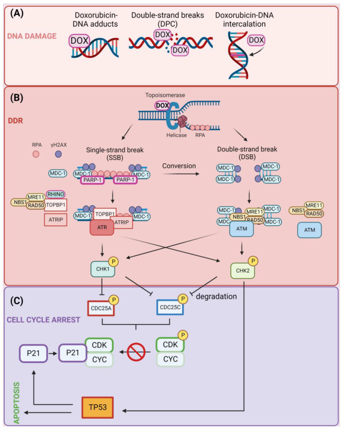
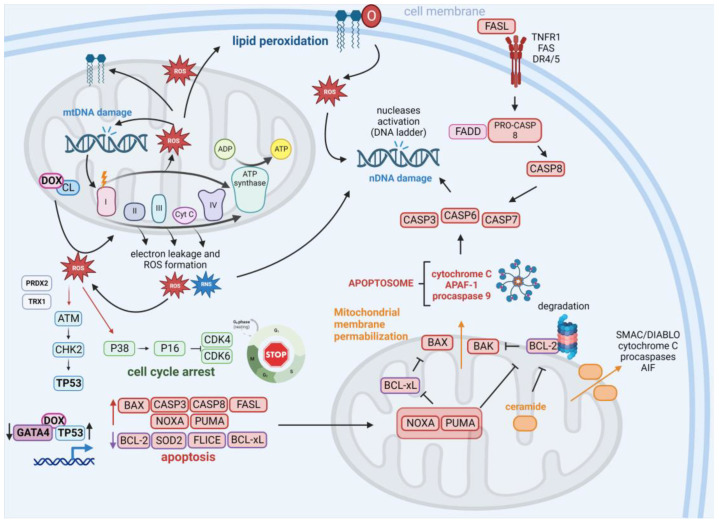
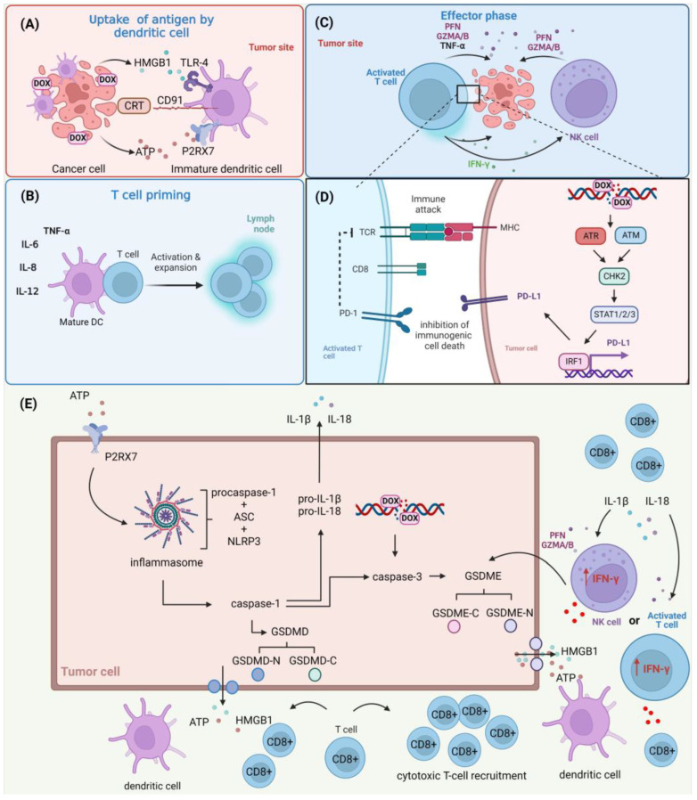
Doxorubicin (DOX) constitutes the major constituent of anti-cancer treatment regimens currently in clinical use. However, the precise mechanisms of DOX's action are not fully understood. Emerging evidence points to the pleiotropic anticancer activity of DOX, including its contribution to DNA damage, reactive oxygen species (ROS) production, apoptosis, senescence, autophagy, ferroptosis, and pyroptosis induction, as well as its immunomodulatory role. This review aims to collect information on the anticancer mechanisms of DOX as well as its influence on anti-tumor immune response, providing a rationale behind the importance of DOX in modern cancer therapy.

Keywords: DNA damage response (DDR); adriamycin; apoptosis; doxorubicin; immunotherapy.


Conflict of interest statement

The authors declare no conflict of interest.

Figures

Figure 1
Figure 1
(A) Induction of DNA damage following treatment of cells with doxorubicin (DOX). DOX induces DNA damage through three main mechanisms: the formation of DNA adducts, single-strand break (SSB) induction, and double-strand break (DSB) induction, in which DNA strands remain bound to trapped topoisomerase enzymes through DNA–protein crosslinks (DPCs) and intercalation of DOX in the DNA molecule. (B) Activation of DNA damage response (DDR) pathway. Trapping of topoisomerase enzymes by DOX induces the formation of either SSBs or DSBs. SSBs are detected by poly [ADP-ribose] polymerase 1 (PARP-1), and the ssDNA is covered with replication protein A (RPA). This results in the recruitment of a variety of factors including DNA topoisomerase 2-binding protein 1 (TOPBP-1), RAD9, RAD1, HUS1 interacting nuclear orphan (RHINO), ATR-interacting protein (ATRIP), MRE11–RAD50–NBS1 complexes, and serine/threonine-protein kinase ATR that contribute to the formation of γH2AX protein foci at the sites of damage. DSBs are detected by MRN complexes, which recruit a mediator of DNA damage checkpoint protein 1 (MDC-1) and amplification of the foci formation by ataxia telangiectasia mutated (A-T mutated) (ATM) kinase. Consequently, ATR and ATM kinases phosphorylate checkpoint kinases CHK1 and CHK2. (C) Induction of apoptosis or cell-cycle arrest by DOX. The CHK1/2-mediated phosphorylation of CDC25A/C phosphatases leads to their degradation and as a consequence no dephosphorylation of CDK-cyclin complexes that prevent cell-cycle progression. Alternatively, CHK2 phosphorylates and activates cellular tumor antigen p53 (TP53) transcription factor leading to up-regulation of P21, which binds unphosphorylated (active) CDK–cyclin complexes and contributes to cell-cycle arrest. Created with BioRender.com accessed on 1 January 2023.
Figure 2
Figure 2
Contribution of DOX to ROS induction and apoptosis. The interaction of DOX with cardiolipin (CL) leads to the elevation of reactive oxygen species (ROS) and reactive nitrogen species (RNS) levels, which contribute to DNA damage of nuclear (nDNA) and mitochondrial DNA (mtDNA). DNA damage and mutations in mtDNA encoding proteins of the electron transport chain (ETC) lead to the production of defective ETC components that increase electron leakage and ROS/RNS generation. Alternatively, ROS trigger lipid peroxidation, enhancing ROS pools in the cells. ROS may trigger the activation of the ATM-CHK2-TP53 signaling independently of DNA damage through ROS-mediated ataxia telangiectasia mutated A-T mutated (ATM) dimer formation through modification of enzyme cysteine residues by peroxiredoxin-2 (PRDX2) and thioredoxin-1 (TRX1). Through the P38 protein, ROS may activate the P16 protein, which works as an inhibitor of cyclin-dependent kinases 4/6 (CDK4/CDK6). DOX increases the expression of cellular tumor antigen p53 (TP53) and down-regulates the expression of GATA binding protein 4 (GATA4), which influences the expression of genes under the control of these transcription factors. Consequently, an increase in multiple proapoptotic proteins (including BAX, caspase 3/8 (CASP3/8), FAS antigen ligand (FASL), phorbol-12-myristate-13-acetate-induced protein 1 (NOXA), and isoform 1 of Bcl-2-binding component 3, isoforms 1/2 (PUMA)) is observed in addition to a decrease in antiapoptotic factors (such as cyclin D1, B-cell lymphoma-extra large (BCL-xL), and FLICE). The observed down-regulation of BCL-xL and apoptosis regulator Bcl-2 (BCL-2) activity can be attributed to the inhibitory activity of NOXA and PUMA and the antiapoptotic protein proteasomal degradation. Alternatively, activation of ceramide signaling together with mitochondrial pore formation (through BAX and BAK proteins) and the loss of mitochondrial integrity confer the release of proapoptotic factors including cytochrome c, apoptotic protease-activating factor 1 (APAF1), and procaspase-9 (leading to apoptosome formation), the secondary mitochondria-derived activator of caspases (SMAC)/direct inhibitor of apoptosis-binding protein with low pI (DIABLO), apoptosis-inducing factor 1, mitochondrial (AIF), and pro-caspases. Apoptosome activity contributes to the activation of effector caspases (CASP3, -6, and -7) that execute the final phase of apoptosis and lead to the activation of nucleases that cleave nDNA (leading to DNA laddering observed following treatment of cells with DOX). DOX also induces the extrinsic apoptosis pathway via up-regulation of FASL, its interaction with the death receptors tumor necrosis factor receptor 1 (TNFR1), FAS, and death receptor 5 (DR5), and activation of CASP8 and effector caspases. Created with BioRender.com accessed on 1 January 2023.
Figure 3
Figure 3
The role of DOX in the anti-tumor immune response. (A) DOX induces the apoptotic cell death of cancer cells, contributing to the exposition or release of ATP, calreticulin (CRT), and high mobility group box protein B1 (HMGB1) of cancer cells and promoting their recognition by dendritic cells (DCs) through respective receptors (P2X purinoceptor 7 (P2RX7), CD91, and toll-like receptor 4 (TLR-4)) present on their cellular surface. (B) These events allow the maturation of DCs and the release of immune-stimulating factors such as interleukins (ILs) and tumor-necrosis factor α (TNF-α). These events promote the activation and expansion of T cells. (C) Activated T cells and natural killer (NK) cells recruited to the tumor microenvironment release perforins (PFNs), granzyme A/B (GZMA/B), and interferon γ (IFN-γ), leading to cancer-cell death. (D) DOX induces double-strand break (DSB) formation, contributing to the activation of the ATR/ATM/CHK2 pathway and promoting STAT1/2/3/IRF1-mediated programmed cell death 1 ligand 1 (PD-L1) up-regulation. This confers to the inhibition of immunogenic cell death. (E) ATP released by dying cancer cells activates P2RX7 expressed on surrounding tumor cells, contributing to the formation of the inflammasome (consisting of adapter protein apoptosis-associated speck-like protein containing a CARD (ASC), NACHT, LRR, and PYD domains-containing protein 3 (NLRP3) and procaspase-1) and activation of caspase-1, which converts pro-IL-1β and pro-IL-18 to active forms (IL-1β, IL-18), activates effector caspase-3, and cleaves gasdermin-D/E (GSDMD/E) to the C-terminal moiety (GSDMD/E-C) and promotes the release of the N-terminal moiety (GSDMD/E-N) that forms pores, triggering pyroptosis. This contributes to the release of a multitude of intracellular factors including ATP, HMGB1, IL-1β, and IL-8 to the extracellular space. Alternatively, the cleavage of GSDMD/E can be triggered by GZNMA/B and PFNs released by NK cells and activated T cells recruited to the tumor microenvironment. Created with BioRender.com accessed on 1 January 2023.

References

    1. Mohseni M., Samadi N., Ghanbari P., Yousefi B., Tabasinezhad M., Sharifi S., Nazemiyeh H. Co-Treatment by Docetaxel and Vinblastine Breaks down P-Glycoprotein Mediated Chemo-Resistance. Iran. J. Basic Med. Sci. 2016;19:300–309. - PMC - PubMed
    1. Dallavalle S., Dobričić V., Lazzarato L., Gazzano E., Machuqueiro M., Pajeva I., Tsakovska I., Zidar N., Fruttero R. Improvement of Conventional Anti-Cancer Drugs as New Tools against Multidrug Resistant Tumors. Drug Resist. Updat. 2020;50:100682. doi: 10.1016/j.drup.2020.100682. - DOI - PubMed
    1. Bukowski K., Kciuk M., Kontek R. Mechanisms of Multidrug Resistance in Cancer Chemotherapy. Int. J. Mol. Sci. 2020;21:3233. doi: 10.3390/ijms21093233. - DOI - PMC - PubMed
    1. Carvalho C., Santos R.X., Cardoso S., Correia S., Oliveira P.J., Santos M.S., Moreira P.I. Doxorubicin: The Good, the Bad and the Ugly Effect. Curr. Med. Chem. 2009;16:3267–3285. doi: 10.2174/092986709788803312. - DOI - PubMed
    1. Rawat P.S., Jaiswal A., Khurana A., Bhatti J.S., Navik U. Doxorubicin-Induced Cardiotoxicity: An Update on the Molecular Mechanism and Novel Therapeutic Strategies for Effective Management. Biomed. Pharmacother. 2021;139:111708. doi: 10.1016/j.biopha.2021.111708. - DOI - PubMed