

Imaging and Histopathological Analysis of Microvascular Angiogenesis in Photodynamic Therapy for Oral Cancer
- PMID: 36831454
- PMCID: PMC9954751
- DOI: 10.3390/cancers15041110
Imaging and Histopathological Analysis of Microvascular Angiogenesis in Photodynamic Therapy for Oral Cancer
Abstract
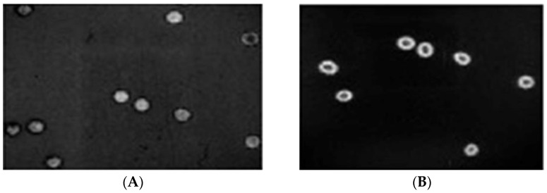
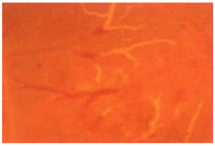
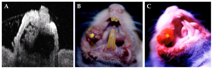
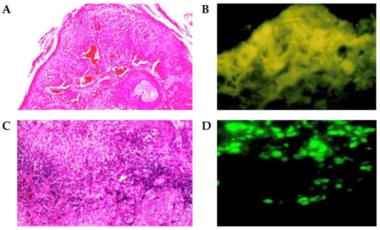
The objective of this study is to use imaging and histopathological analysis to characterize and monitor microvascular responses to photodynamic therapy (PDT). In vivo chicken chorioallantoic membranes (CAMs) and a stimulated malignant oral lesions animal model were used to determine the blood flow and the biological activities of Photofrin® (2.5 mg/kg) exposed to different laser power densities at 630 nm. The vascular changes, the velocity of the blood flow, the speckle flow index (SFI) of fluorescence changes, and ultrastructure damage in the microvasculature before and after PDT were recorded. The subcellular localization of Photofrin® revealed satisfactory uptake throughout the cytoplasm of human red blood cells at 10 s and 20 s before PDT. The mean blood-flow velocities of the veins and arteries were 500 ± 40 and 1500 ± 100 μm/s, respectively. A significant decrease in the velocities of the blood flow in the veins and arteries was detected in the CAM model after PDT. The veins and arteries of CAMs, exposed to the power densities of 80, 100, and 120 mW/cm2, had average blood-flow velocities of 100 ± 20, 60 ± 10, and 0 μm/s and 300 ± 50, 150 ± 30, and 0 μm/s, respectively. In the stimulated malignant oral lesions animal model, the treated tumors exhibited hemorrhage and red blood cell extravasation after PDT. The oxyhemoglobin and total hemoglobin levels decreased, which resulted in a decrease in tissue oxygen saturation, while the deoxyhemoglobin levels increased. PDT using Photofrin® has the ability to cause the destruction of the targeted microvasculature under nonthermal mechanisms selectively.
Keywords: chicken chorioallantoic membrane; photodynamic therapy; stimulated malignant oral lesions.
Conflict of interest statement
The authors declare no conflict of interest.
Figures
Similar articles
-
In vitro and in vivo photosensitizing capabilities of 5-ALA versus photofrin in vascular endothelial cells.Lasers Surg Med. 1999;24(3):178-86. doi: 10.1002/(sici)1096-9101(1999)24:3<178::aid-lsm2>3.0.co;2-w. Lasers Surg Med. 1999. PMID: 10229148
-
Microvascular effects of Photofrin(®)-induced photodynamic therapy.Photodiagnosis Photodyn Ther. 2007 Jun;4(2):95-9. doi: 10.1016/j.pdpdt.2007.03.003. Epub 2007 May 7. Photodiagnosis Photodyn Ther. 2007. PMID: 25047341
-
Photodynamic therapy for premalignant lesions in DMBA-treated hamsters: a preliminary study.J Oral Maxillofac Surg. 1997 Apr;55(4):376-81; discussion 381-2. doi: 10.1016/s0278-2391(97)90130-0. J Oral Maxillofac Surg. 1997. PMID: 9120701
-
The Chicken Embryo Chorioallantoic Membrane as an In Vivo Model for Photodynamic Therapy.Methods Mol Biol. 2022;2451:107-125. doi: 10.1007/978-1-0716-2099-1_9. Methods Mol Biol. 2022. PMID: 35505014 Review.
-
Photodynamic therapy and cancer of the esophagus.Semin Oncol. 1994 Dec;21(6 Suppl 15):20-3. Semin Oncol. 1994. PMID: 7992103 Review.
Cited by
-
Photodynamic Therapy for Oral Squamous Cell Carcinoma: Current Status, Challenges, and Prospects.Int J Nanomedicine. 2024 Oct 22;19:10699-10710. doi: 10.2147/IJN.S481901. eCollection 2024. Int J Nanomedicine. 2024. PMID: 39464676 Free PMC article. Review.
References
-
- Spikes J.D. The historical development of ideas on applications of photosensitized reactions in the health sciences. In: Benasson R.V., Jori G., Land E.J., Truscott T.G., editors. Primary Photo-Processes in Biology and Medicine. Plenum Press; New York, NY, USA: 1985. pp. 209–227.
-
- Chang C.J., Sun C.H., Liaw L.H.L., Nelson J.S., Berns M.W. In vitro and in vivo photosensitizing capabilities of 5-ALA compared to Photofrin® in vascular endothelial cells. Lasers Surg. Med. 1999;24:178–186. - PubMed
LinkOut - more resources
Full Text Sources
