Review
. 2023 Feb 19;15(4):1316.
doi: 10.3390/cancers15041316.

Downstream Targets of VHL/HIF-α Signaling in Renal Clear Cell Carcinoma Progression: Mechanisms and Therapeutic Relevance

Affiliations
Review

Downstream Targets of VHL/HIF-α Signaling in Renal Clear Cell Carcinoma Progression: Mechanisms and Therapeutic Relevance

Sonia Mazumder et al. Cancers (Basel). .

Abstract

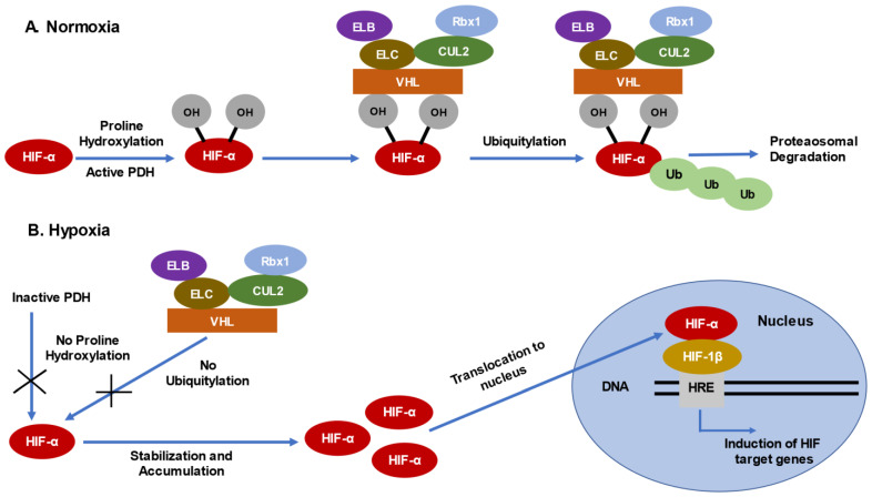
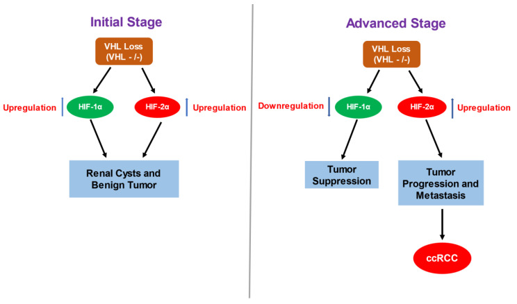
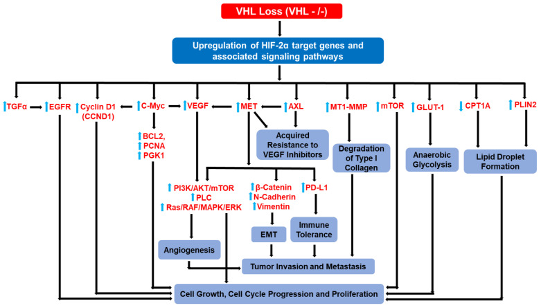
The clear cell variant of renal cell carcinoma (ccRCC) is the most common renal epithelial malignancy and responsible for most of the deaths from kidney cancer. Patients carrying inactivating mutations in the Von Hippel-Lindau (VHL) gene have an increased proclivity to develop several types of tumors including ccRCC. Normally, the Hypoxia Inducible Factor alpha (HIF-α) subunits of the HIF heterodimeric transcription factor complex are regulated by oxygen-dependent prolyl-hydroxylation, VHL-mediated ubiquitination and proteasomal degradation. Loss of pVHL function results in elevated levels of HIF-α due to increased stability, leading to RCC progression. While HIF-1α acts as a tumor suppressor, HIF-2α promotes oncogenic potential by driving tumor progression and metastasis through activation of hypoxia-sensitive signaling pathways and overexpression of HIF-2α target genes. One strategy to suppress ccRCC aggressiveness is directed at inhibition of HIF-2α and the associated molecular pathways leading to cell proliferation, angiogenesis, and metastasis. Indeed, clinical and pre-clinical data demonstrated the effectiveness of HIF-2α targeted therapy in attenuating ccRCC progression. This review focuses on the signaling pathways and the involved genes (cyclin D, c-Myc, VEGF-a, EGFR, TGF-α, GLUT-1) that confer oncogenic potential downstream of the VHL-HIF-2α signaling axis in ccRCC. Discussed as well are current treatment options (including receptor tyrosine kinase inhibitors such as sunitinib), the medical challenges (high prevalence of metastasis at the time of diagnosis, refractory nature of advanced disease to current treatment options), scientific challenges and future directions.

Keywords: EGFR; HIF-α; MET; VHL; ccRCC.


Conflict of interest statement

The authors declare no conflict of interest.

Figures

Figure 1
Figure 1
Regulation of HIF-α via VHL-dependent ubiquitylation and proteasomal degradation. (A) Alpha subunits of the Hypoxia Inducible Factors (HIFs) are constitutively expressed but are tightly regulated. Under normal oxygen availability, HIF-α subunits are hydroxylated at specific proline residues via the activity of Proline Hydroxylases (PDHs), which target HIF-α for degradation via VHL- dependent recognition and ubiquitination. (B) Under low oxygen availability, however, PDH-dependent hydroxylation of proline residues in HIF-α is absent; as a result, recognition via VHL and subsequent HIF-α degradation is inhibited. Consequently, HIF-α subunits are stabilized, accumulate, and translocate to the nucleus, forming heterodimeric complexes with HIF-1β subunits. This complex interacts with the (HIF Response Element) HRE in DNA to induce the expression of HIF target genes that regulate angiogenesis, cell growth, cell cycle progression, cell proliferation, glycolysis, and apoptosis to maintain cell growth and survival under hypoxic conditions.
Figure 2
Figure 2
Contrasting Role of HIF-1α and HIF-2α in ccRCC progression. Loss of VHL leads to the upregulation of HIF-α isoforms: HIF-1α and HIF-2α, irrespective of oxygen availability in cell, which in turn contributes to the formation of initial renal cysts and tumors. Interestingly, only the HIF-1α isoform is expressed in normal tubular cells and the initiation of HIF-2α expression upon VHL loss, acts as an initial trigger for transformation of healthy tubular cells. Overtime, the expression of HIF-2α prevails over HIF-1α expression, which is associated with tumor growth, progression and metastasis. Furthermore, HIF-1α attenuates expression of several pro-tumorigenic genes which otherwise are stimulated by HIF-2α suppressing the malignant phenotype.
Figure 3
Figure 3
HIF-2α-induced genetic responses and the associated oncogenic hierarchical events leading to ccRCC progression. HIF-2α induces the expression of multiple target genes (e.g., EGFR, CCND1, c-Myc, MET, VEGF, mTOR, GLUT1, CPTA1). Such gene reprogramming events promote angiogenesis, cell proliferation/growth, cell cycle progression, EMT, tumor migration/invasion, anaerobic glycolysis, lipid accumulation, immune tolerance, and drug resistance, which collectively lead to advanced ccRCC. Various functional interplay among target proteins and downstream pathogenic consequences are depicted above. VEGF-mediated tumor angiogenesis, for example, can promote tumor invasion and growth. Similarly, metabolic reprogramming (e.g., glycolysis induction) initiated by Glut1 can sustain cancer growth.

Similar articles

Cited by

References

    1. Cho H., Kaelin W.G. Targeting HIF2 in clear cell renal cell carcinoma. Cold Spring Harb. Symp. Quant. Biol. 2016;81:113–121. doi: 10.1101/sqb.2016.81.030833. - DOI - PubMed
    1. D’Avella C., Abbosh P., Pal S.K., Geynisman D.M. Mutations in renal cell carcinoma. Urol. Oncol. Semin. Orig. Investig. 2020;38:763–773. doi: 10.1016/j.urolonc.2018.10.027. - DOI - PubMed
    1. Frew I.J., Moch H. A clearer view of the molecular complexity of clear cell renal cell carcinoma. Annu. Rev. Pathol. Mech. Dis. 2015;10:263–289. doi: 10.1146/annurev-pathol-012414-040306. - DOI - PubMed
    1. Batavia A.A., Schraml P., Moch H. Clear cell renal cell carcinoma with wild-type von Hippel-Lindau gene: A non-existent or new tumour entity? Histopathology. 2019;74:60–67. doi: 10.1111/his.13749. - DOI - PubMed
    1. Ericsson J.L., Seljelid R., Orrenius S. Comparative light and electron microscopic observations of the cytoplasmic matrix in renal carcinomas. Virchows Arch. Pathol. Anat. Physiol. Klin. Med. 1966;341:204–223. doi: 10.1007/BF00961071. - DOI - PubMed