Review
. 2023 Feb 19;15(4):1320.
doi: 10.3390/cancers15041320.

Targeted Therapy and Mechanisms of Drug Resistance in Breast Cancer

Affiliations
Review

Targeted Therapy and Mechanisms of Drug Resistance in Breast Cancer

Briana Kinnel et al. Cancers (Basel). .

Abstract

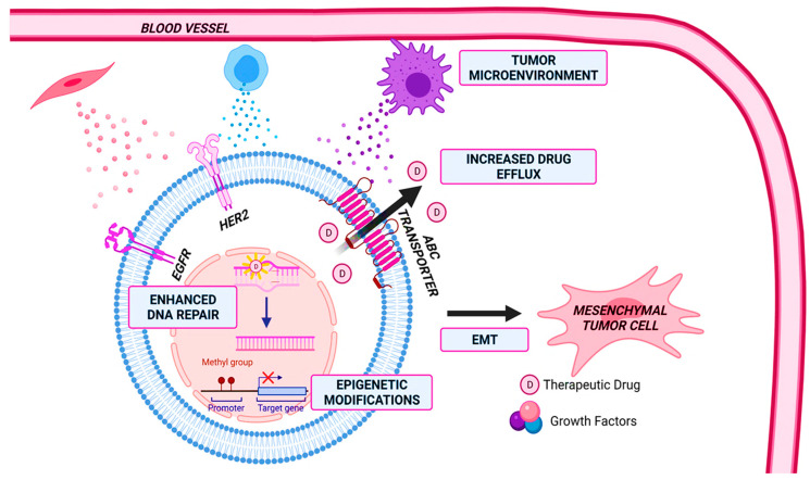
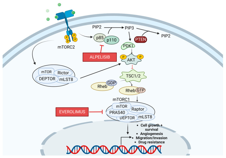
Breast cancer is the most common cause of cancer-related death in women worldwide. Multidrug resistance (MDR) has been a large hurdle in reducing BC death rates. The drug resistance mechanisms include increased drug efflux, enhanced DNA repair, senescence escape, epigenetic alterations, tumor heterogeneity, tumor microenvironment (TME), and the epithelial-to-mesenchymal transition (EMT), which make it challenging to overcome. This review aims to explain the mechanisms of resistance in BC further, identify viable drug targets, and elucidate how those targets relate to the progression of BC and drug resistance.

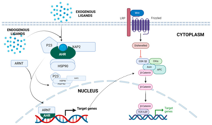
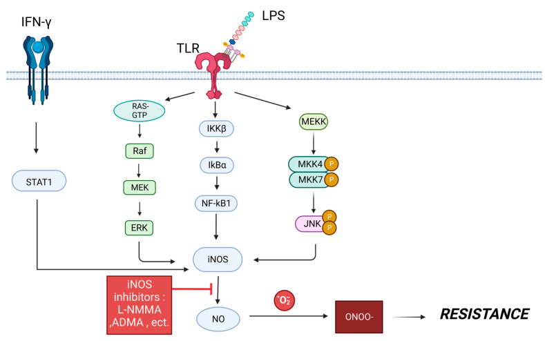
Keywords: CDK4/6; HER2; MDR; PTK6; Wnt/β-catenin; aryl hydrocarbon receptor; drug resistance; iNOS.


Conflict of interest statement

The authors declare that they have no competing interests.

Figures

Figure 1
Figure 1
Mechanisms of drug resistance in breast cancer. These mechanisms include changes in the tumor microenvironment, enhanced DNA repair, epigenetic modifications, epithelial-to-mesenchymal transition (EMT), and increased drug efflux. Figure created with Biorender.com.
Figure 2
Figure 2
The Aryl Hydrocarbon Receptor (AhR) pathway. When inactive, the AhR is located in the cytoplasm and forms a complex with XAP2, also known as AIP, two HSP90 molecules, and p23. After ligand interaction, AhR translocates to the nucleus, interacting with ARNT to create a heterodimer. The heterodimer binds to the XRE and coregulators, leading to the transcription of target genes. AhR crosstalk with the Wnt pathway is also displayed. Figure created with Biorender.com.
Figure 3
Figure 3
Signaling pathways that induce inducible nitric oxide synthase (iNOS) production, leading to nitrous oxide (NO) production. These pathways include the signal transducer and activator of transcription 1 (STAT1), Ras/Raf/MAPK, an inhibitor of nuclear factor kappa-B kinase (IKKβ), and c-Jun N-terminal kinase (JNK) pathways. NO reacts with oxygen free radicals to produce peroxynitrite, which can lead to drug resistance. NO production could potentially be reduced using iNOS inhibitors such as NG-monomethyl-L-arginine acetate (L-NMMA) and asymmetric dimethylarginine (ADMA). Created with BioRender.com.
Figure 4
Figure 4
Diagram of the p85/p110 (PI3K) signaling pathway. PI3K leads to the formation of the second messenger, PIP3. PDK1 and AKT are then recruited to the membrane. PDK1 and mTORC2 phosphorylate AKT, fully activating it. AKT then phosphorylates TSC, which is a negative regulator of mTORC1. Alpelisib is a PI3K inhibitor, and Everolimus is an mTOR inhibitor. Created with BioRender.com.
Figure 5
Figure 5
The effects of PARP binding after single-stranded DNA breaks. The binding of PARP1 helps detect the single-stranded break (SSB) and promotes the base excision repair (BER) mechanism for DNA repair. PARP must be released from the DNA for the repair to occur. When treated with PARP inhibitors (PARPi), PARP trapping occurs, leading to double-stranded breaks (DSB). When homologous recombination (HR) occurs, the DNA is repaired, and cell survival occurs. Created with BioRender.com.
Figure 6
Figure 6
A diagram of PTK6 signaling. PTK6 activates several signaling pathways, such as JAK/STAT, PI3K, and MAPK, which enhance cell proliferation, invasion/migration, and resistance. Created with BioRender.com.
Figure 7
Figure 7
A diagram of how the binding of CDK4/6 relates to cell cycle progression. RAS signaling and estrogen receptor (ER) activation lead to the production of cyclin D. The binding of Cyclin D to CDK4/6 helps push the cell cycle from the G1 to S phase via Rb phosphorylation. The nodes at which current therapies target the pathway are included as well. Created with BioRender.com.
Figure 8
Figure 8
A diagram of the Wnt signaling pathway. The binding of Wnt to its receptors Frizzled and LRP5/6 inhibits the degradation of β-catenin, which regulates the expression of many genes. Receptor activation leads to the recruitment of the disheveled protein (Dvl). Dvl is then activated via phosphorylation, inducing the dissociation of GSK-3β from axin and causing GSK-3β to be deactivated. The deactivation of GSK-3β inhibits the degradation of β-catenin, allowing it to translocate into the nucleus. The nodes at which current therapies target the pathway are included as well. Created with BioRender.com.
Figure 9
Figure 9
EGFR, HER2, HER3, HER4 signaling pathways. Created with Biorender.com.

References

    1. Sung H., Ferlay J., Siegel R.L., Laversanne M., Soerjomataram I., Jemal A., Bray F. Global Cancer Statistics 2020: GLOBOCAN Estimates of Incidence and Mortality Worldwide for 36 Cancers in 185 Countries. CA Cancer J. Clin. 2021;71:209–249. doi: 10.3322/caac.21660. - DOI - PubMed
    1. Zhao H. The prognosis of invasive ductal carcinoma, lobular carcinoma and mixed ductal and lobular carcinoma according to molecular subtypes of the breast. Breast Cancer. 2020;28:187–195. doi: 10.1007/s12282-020-01146-4. - DOI - PubMed
    1. Łukasiewicz S., Czeczelewski M., Forma A., Baj J., Sitarz R., Stanisławek A. Breast Cancer-Epidemiology, Risk Factors, Classification, Prognostic Markers, and Current Treatment Strategies-An Updated Review. Cancers. 2021;13:4287. doi: 10.3390/cancers13174287. - DOI - PMC - PubMed
    1. Johnson K.S., Conant E.F., Soo M.S. Molecular Subtypes of Breast Cancer: A Review for Breast Radiologists. J. Breast Imaging. 2020;3:12–24. doi: 10.1093/jbi/wbaa110. - DOI - PubMed
    1. Waks A.G., Winer E.P. Breast Cancer Treatment: A Review. JAMA. 2019;321:288–300. doi: 10.1001/jama.2018.19323. - DOI - PubMed