. 2023 Jan 18;13(2):164.
doi: 10.3390/brainsci13020164.

Targeting the Tumor Immune Microenvironment Could Become a Potential Therapeutic Modality for Aggressive Pituitary Adenoma

Affiliations

Targeting the Tumor Immune Microenvironment Could Become a Potential Therapeutic Modality for Aggressive Pituitary Adenoma

Zuocheng Yang et al. Brain Sci. .

Abstract

Object: This study aimed to explore the relationship between the aggressiveness and immune cell infiltration in pituitary adenoma (PA) and to provide the basis for immuno-targeting therapies.

Methods: One hundred and three patients with PA who underwent surgery at a single institution were retrospectively identified. The infiltration of macrophages and T-lymphocytes was quantitatively assessed.

Results: The number of CD68+ macrophages was positively correlated with Knosp (p = 0.003) and MMP-9 expression grades (p = 0.00). The infiltration of CD163+ macrophages differed among Knosp (p = 0.022) and MMP-9 grades (p = 0.04). CD8+ tumor-infiltrating lymphocytes (TILs) were also positively associated with Knosp (p = 0.002) and MMP-9 grades (p = 0.01). Interestingly, MGMT expression was positively correlated with MMP-9 staining extent (p = 0.000). The quantities of CD8+ TILs (p = 0.016), CD68+ macrophages (p = 0.000), and CD163+ macrophages (p = 0.043) were negatively associated with MGMT expression levels. The number of CD68+ macrophages in the PD-L1 negative group was significantly more than that in the PD-L1 positive group (p = 0.01). The rate of PD-L1 positivity was positively correlated with the Ki-67 index (p = 0.046) and p53 expression (p = 0.029).

Conclusion: Targeted therapy for macrophages and CD8+ TILs could be a helpful treatment in the future for aggressive PA. Anti-PD-L1 therapy may better respond to PAs with higher Ki-67 and p53 expression and more infiltrating CD68+ macrophages. Multiple treatment modalities, especially combined with immunotherapy could become a novel therapeutic strategy for aggressive PA.

Keywords: CD8+ TILs; aggressive pituitary adenoma; combined immunotherapy; immune microenvironment; macrophage; temozolomide.


Conflict of interest statement

The authors declare that they have no conflict of interest.

Figures

Figure 1
Figure 1
Immunohistochemistry of tissue microarray for immune cells in pituitary adenoma (PA). The different distribution of CD8+ TILs between the non-aggressive PA and aggressive PA at ×400 magnification (a) and the Knosp grades (b); the different distribution of CD68+ macrophages surrounding the vessels between the non-aggressive PA and aggressive PA at ×400 magnification (c) and the Knosp grades (d); the different distribution of the density of infiltrating CD163+ macrophages between the non- aggressive PA and aggressive PA at ×400 magnification (e) and the Knosp grades (f).
Figure 2
Figure 2
Correlation heatmap (Spearman correlation) of immune cells and pro-invasive factors. Only statistically significant differences are displayed.
Figure 3
Figure 3
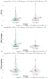
Immunohistochemical studies of O6-methylguanine-DNA methyltransferase (MGMT), PD-L1 and metalloproteinase (MMP)-9 in PA. The different MGMT expression between non-aggressive PA and aggressive PA at ×200 magnification (a) and the Knosp grades (b); the different PD-L1 expression between non-aggressive PA and aggressive PA at ×200 magnification (c) and the Knosp grades (d); the different MMP-9 expression between non-aggressive PA and aggressive PA at ×200 magnification (e) and the Knosp grades (f).
Figure 4
Figure 4
Unpaired t-test performed between the PD-L1 positive and PD-L1 negative groups in CD8+ TILs (a); the infiltration of CD68+ macrophages difference between PD-L1 positive and PD-L1 negative groups were calculated with an unpaired t-test (b); Student’s t-test between the PD-L1 positive and PD-L1 negative groups in CD168+ macrophages (c).
Figure 5
Figure 5
Proportions of positive PD-L1 between null cell and non-null cell groups are compared using the chi-squared test.
Figure 6
Figure 6
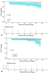
Kaplan–Meier PFS curves. PFS curve and PFS stratification based on complete resection (CR) and incomplete resection (IR) (a); PFS differed significantly between the MMP-9 positive and MMP-9 negative groups (b).

Similar articles

Cited by

References

    1. Melmed S. Pituitary-Tumor Endocrinopathies. N. Engl. J. Med. 2020;382:937–950. doi: 10.1056/NEJMra1810772. - DOI - PubMed
    1. Molitch M.E. Diagnosis and Treatment of Pituitary Adenomas: A Review. JAMA. 2017;317:516–524. doi: 10.1001/jama.2016.19699. - DOI - PubMed
    1. Heaney A. Management of aggressive pituitary adenomas and pituitary carcinomas. J. Neurooncol. 2014;117:459–468. doi: 10.1007/s11060-014-1413-6. - DOI - PubMed
    1. Nomikos P., Buchfelder M., Fahlbusch R. The outcome of surgery in 668 patients with acromegaly using current criteria of biochemical ‘cure’. Eur. J. Endocrinol. 2005;152:379–387. - PubMed
    1. Rhee I. Diverse macrophages polarization in tumor microenvironment. Arch. Pharm. Res. 2016;39:1588–1596. doi: 10.1007/s12272-016-0820-y. - DOI - PubMed