. 2023 Feb 1;13(2):253.
doi: 10.3390/brainsci13020253.

Auricular Vagus Nerve Stimulation Improves Visceral Hypersensitivity and Gastric Motility and Depression-like Behaviors via Vago-Vagal Pathway in a Rat Model of Functional Dyspepsia

Affiliations

Auricular Vagus Nerve Stimulation Improves Visceral Hypersensitivity and Gastric Motility and Depression-like Behaviors via Vago-Vagal Pathway in a Rat Model of Functional Dyspepsia

Liwei Hou et al. Brain Sci. .

Abstract

Transcutaneous auricular vagus nerve stimulation was recently reported to have a therapeutic potential for functional dyspepsia (FD). This study aimed to explore the integrative effects and mechanisms of auricular vagus nerve stimulation (aVNS) in a rodent model of FD.

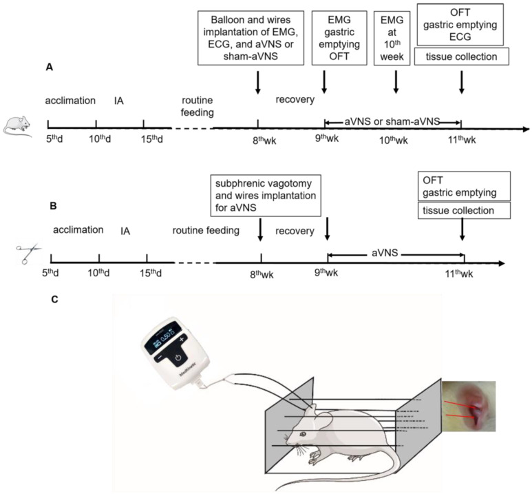
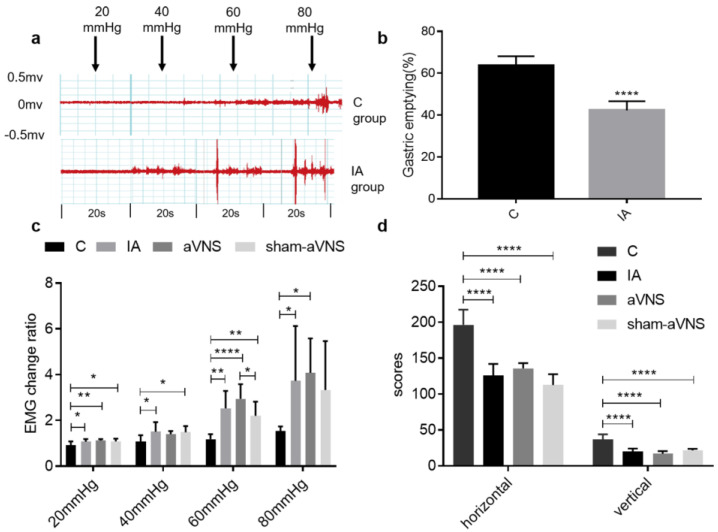
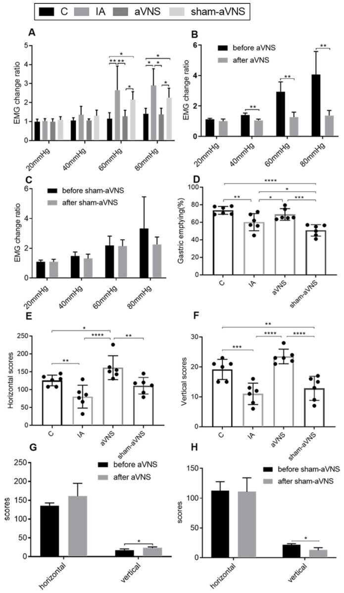
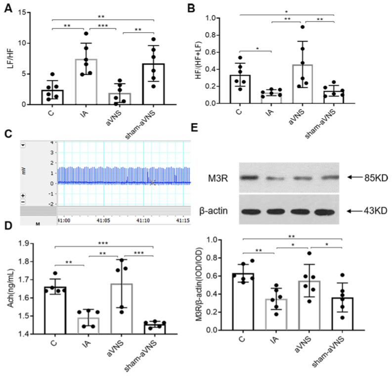
Methods: We evaluated the effects of aVNS on visceral hypersensitivity, gastric motility and open field test (OFT) activity in iodoacetamide (IA)-treated rats. The autonomic function was assessed; blood samples and tissues were collected and analyzed by an enzyme-linked immunosorbent assay and western blot. Vagotomy was performed to investigate the role of vagal efferent nerve.

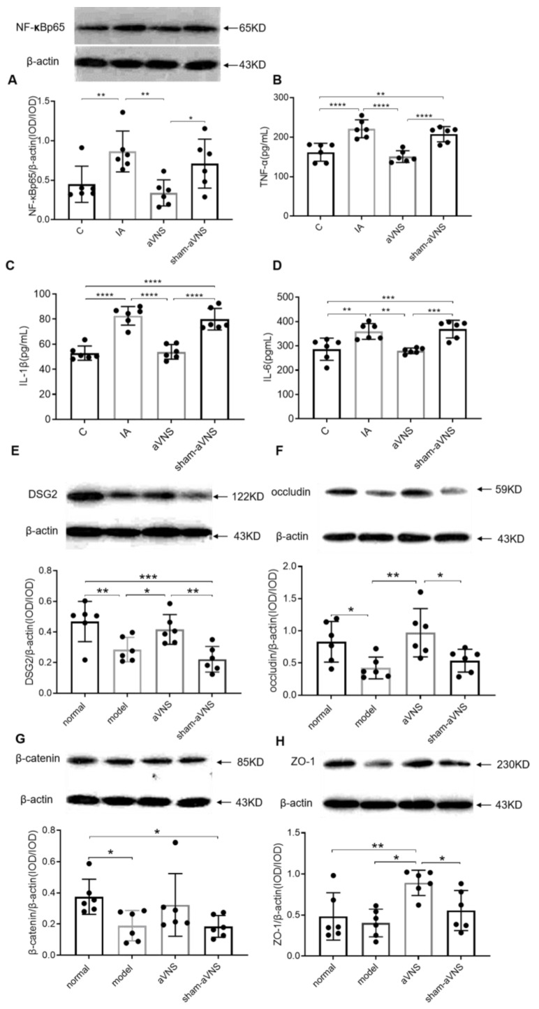
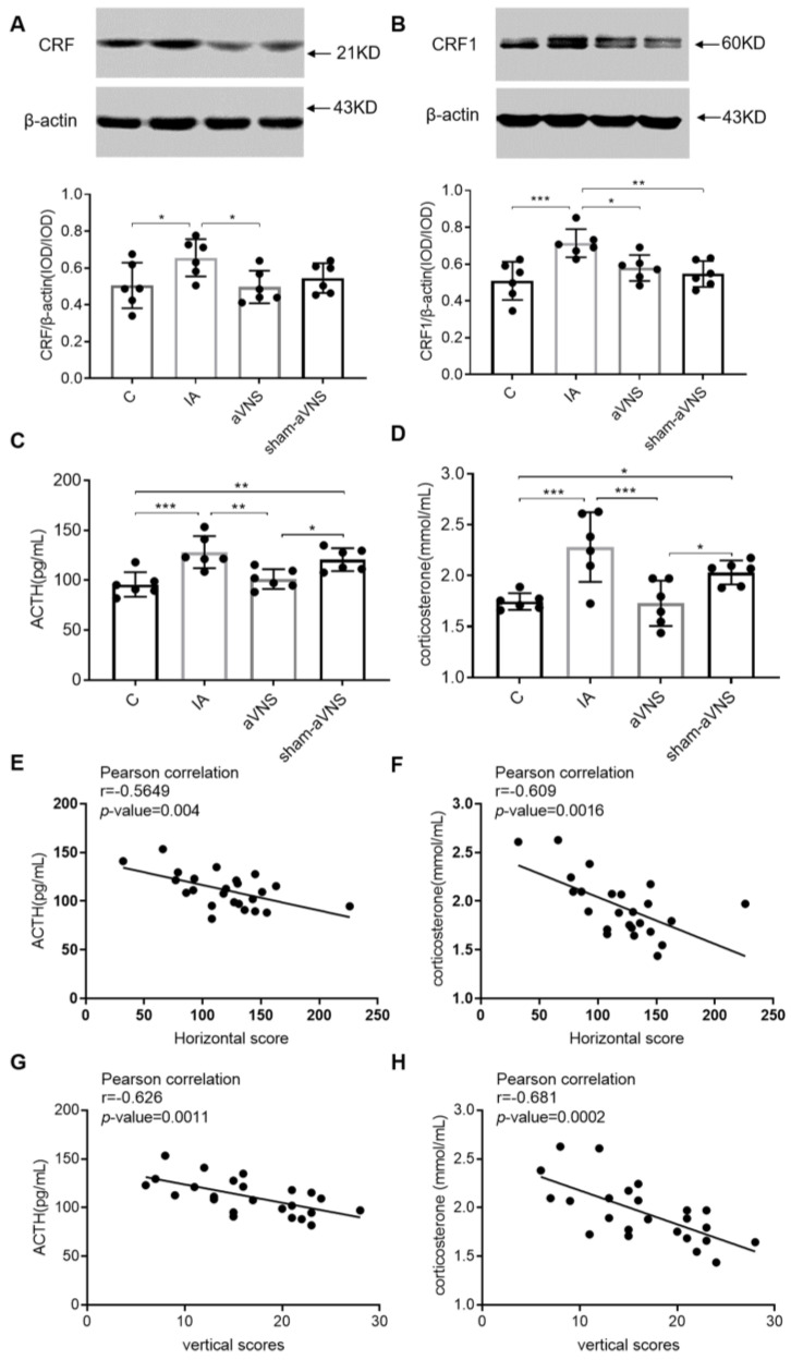
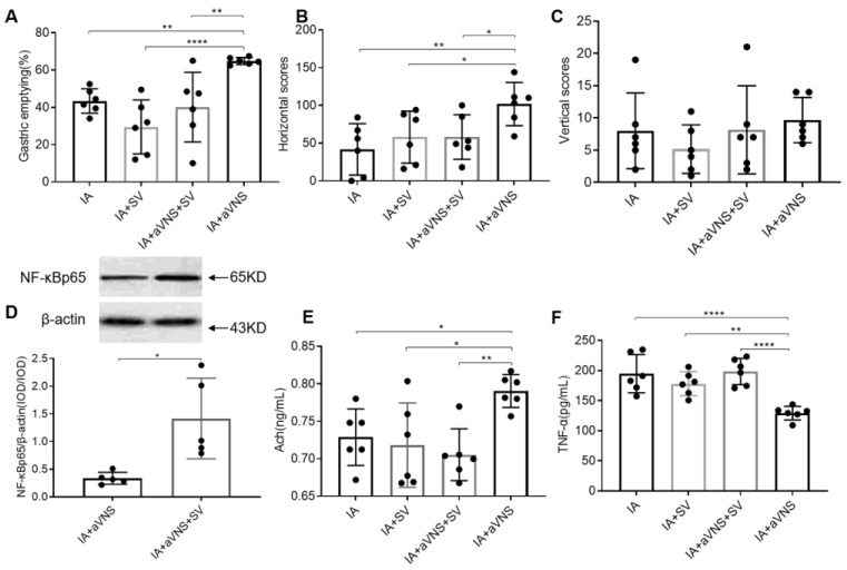
Results: aVNS reduced the electromyography response to gastric distension, improved gastric emptying and increased the horizontal and vertical motion scores of the OFT in IA-treated rats. The sympathovagal ratio was increased in IA-treated rats but normalized with aVNS. The serum cytokines TNF-α, IL-6, IL-1β and NF-κBp65 were increased in IA-treated rats and decreased with aVNS. The hypothalamus-pituitary-adrenal axis was hyperactive in IA-treated rats but inhibited by aVNS. The expression of duodenal desmoglein 2 and occludin were all decreased in IA-treated rats and increased with aVNS but not sham-aVNS. Vagotomy abolished the ameliorating effects of aVNS on gastric emptying, horizontal motions, serum TNF-α and duodenal NF-κBp65.

Conclusion: aVNS improves gastric motility and gastric hypersensitivity probably by anti-inflammatory mechanisms via the vago-vagal pathways. A better understanding on the mechanisms of action involved with aVNS would lead to the optimization of the taVNS methodology and promote taVNS as a non-pharmacological alternative therapy for FD.

Keywords: cholinergic anti-inflammatory pathway; hypothalamic–pituitary-adrenal axis; inflammation; vagotomy; vagus nerve.


Conflict of interest statement

The authors declare no conflict of interest.

Figures

Figure 1
Figure 1
(A) Flowchart of experiment one. All ten-day-old SD rats except for control rats received iodoacetamide (IA) daily for 6 days and housed normally to grow until adulthood of 8 weeks old. Surgical implantations for ECG and EMG were performed at the age of 8 weeks and then recovered for 1 week. Physiological measurements were made at the 9th week and after the 11th week. Wire implantation for aVNS or sham-aVNS were performed at the age of 8 weeks and then recovered for 1 week at each group. The treatment (aVNS or sham-aVNS) was conducted for 2 weeks (9th week to 11th week). Only the EMG was performed after the 9th week and 10th week. (B) Flowchart of experiment two. All ten-day-old SD rats received the same IA treatment. Subphrenic vagotomy were performed at the age of 8 weeks and then recovered for 1 week. aVNS was conducted as experiment one. Physiological measurements were made after the 11th week. (C) aVNS schematic diagram.
Figure 2
Figure 2
Assessment of rat model. (a) Different EMG waveforms changes response to gastric distension (GD) of 20, 40, 60 and 80 mmHg in control rats and IA-treated rats. (b) Gastric emptying results in control rats and IA-treated rats. The index was detected from 12 sacrificed rats after the 9th week (6 rats were randomly picked in control group and IA-treated group, respectively). The independent sample t-test was used. (c) EMG change ratio response to GD in all rats when rat model was induced. (d) The open field test (OFT) results when rat model was induced in all rats. All indexes were tested after the 9th week and before aVNS or sham-aVNS. One-way ANOVA was used, and the LSD method was used for post hoc tests in EMG change ratio and OFT. EMG: electromyography. * p < 0.05, ** p < 0.01, **** p < 0.0001. C: control group; IA: IA-treated group. n = 6 per group. Data were presented as means ± standard deviation.
Figure 3
Figure 3
Effects of the aVNS. (A) EMG change ratio results after one-week intervention in all rats. (B) Comparison results of EMG change ratio before and after aVNS. (C) Comparison results of EMG change ratio before and after sham-aVNS. (D) Gastric emptying after two-week intervention in all rats. (E,F) OFT results after two-week intervention in all rats. (G) Comparison results of horizontal scores before and after aVNS. (H) Comparison results of vertical scores before and after sham-aVNS. A paired t-test was adopted for comparison in (B,C,G,H). One-way ANOVA was used, and the LSD method was used for post hoc tests in (A,D,E,F) (except for 80 mmHg in A Tamhane’s T2(M) method was used). EMG: electromyography. * p < 0.05, ** p < 0.01, *** p < 0.001, **** p < 0.0001. C: control group; IA: IA-treated group. n = 6 per group. Data were presented as means ± standard deviation.
Figure 4
Figure 4
aVNS increased vagal activity, improved sympathovagal imbalance, promoted the level of Ach and its receptor of M3R in gastric tissue. (A) LF/HF results after two weeks of intervention in all rats. (B) HF/(HF + LF) results after two weeks of intervention in all rats. (C) ECG waveform in 15 s. (D) Acetylcholine concentration from gastric tissue in all results. (E) The expression of gastric M3R in all rats. LF: low frequency, represents sympathetic nerve activity. HF: high frequency represents vagal nerve activity. One-way ANOVA was used, and the LSD method was used for post hoc tests for all indexes. * p < 0.05, ** p < 0.01, *** p < 0.001. ECG: electrocardiograph. C: control group; IA: IA-treated group. n = 6 per group. Data were presented as means ± standard deviation.
Figure 5
Figure 5
The anti-inflammatory and mucosal integrity mechanism of the intervention. (A) Level of duodenal NF-κBp65 after two weeks of intervention in all groups. (BD) Release of serum TNF-α, IL-1β and IL-6 after two weeks of intervention in all groups. (EH) Expressions of duodenal l proteins DSG2, occludin, β-catenin and ZO-1. One-way ANOVA was used, and the LSD method was used for post hoc tests for all. * p < 0.05, ** p < 0.01, *** p < 0.001, **** p < 0.0001. NF-κBp65: nuclear factor kappa Bp65.TNF-α: tumor necrosis factorα.IL-1β: interleukin 1β. IL-6: interleukin 6. DSG2: desmoglein2. ZO-1: zonula occluden. C: control group; IA: IA-treated group. n = 6 per group. Data were presented as means ± standard deviation.
Figure 6
Figure 6
The hypothalamic–pituitary–adrenal axis mechanism of the intervention. (A) Expression of hypothalamus CRF in all rats. (B) Expression of amygdaloid nucleus CRF1 in all rats. (C,D) The release of serum ACTH and corticosterone in all rats. One-way ANOVA was used, and the LSD method was used for post hoc tests for all. (EH): Pearson correlation analysis between HPA axis and OFT. * p < 0.05, ** p < 0.01, *** p < 0.001. ACTH: adrenocorticotropic hormone. CRF: corticotropin releasing factor. C: control group; IA: IA-treated group. n = 6 per group. Data were presented as means ± standard deviation.
Figure 7
Figure 7
The effect and mechanism of vagotomy in IA-treated rats. (AC) The gastric emptying and OFT results in all rats. (D) Expression of duodenal NF-κBp65 after two weeks of intervention. (E) The release of gastric Ach in all rats. (F) The serum level of inflammatory cytokines TNF-α in all rats. One-way ANOVA was used, and the LSD method was used for post hoc tests in (AC,E,F). The independent sample t-test was used in (D). IA: IA-treated rats. IA + SV group: IA-treated rats received SV with no intervention. IA + aVNS group: IA-treated rats with 2 weeks of aVNS; IA + aVNS + SV group: IA-treated rats received SV with 2 weeks of aVNS. SV: subphrenic vagotomy. * p < 0.05, ** p < 0.01, **** p < 0.0001. n = 6 per group. Data were presented as means ± standard deviation.

Similar articles

Cited by

References

    1. Ford A.C., Mahadeva S., Carbone M.F., Lacy B.E., Talley N.J. Functional dyspepsia. Lancet. 2020;396:1689–1702. doi: 10.1016/S0140-6736(20)30469-4. - DOI - PubMed
    1. Aziz I., Palsson O.S., Tornblom H., Sperber A.D., Whitehead W.E., Simren M. Epidemiology, clinical characteristics, and associations for symptom-based Rome IV functional dyspepsia in adults in the USA, Canada, and the UK: A cross-sectional population-based study. Lancet Gastroenterol. Hepatol. 2018;3:252–262. doi: 10.1016/S2468-1253(18)30003-7. - DOI - PubMed
    1. Moayyedi P., Lacy B.E., Andrews C.N., Enns R.A., Howden C.W., Vakil N. ACG and CAG Clinical Guideline: Management of Dyspepsia. Am. J. Gastroenterol. 2017;112:988–1013. doi: 10.1038/ajg.2017.154. - DOI - PubMed
    1. Van Den Houte K., Carbone F., Tack J. Postprandial distress syndrome: Stratification and management. Expert Rev. Gastroenterol. Hepatol. 2019;13:37–46. doi: 10.1080/17474124.2019.1543586. - DOI - PubMed
    1. Lacy B.E., Weiser K.T., Kennedy A.T., Crowell M.D., Talley N.J. Functional dyspepsia: The economic impact to patients. Aliment. Pharmacol. Ther. 2013;38:170–177. doi: 10.1111/apt.12355. - DOI - PubMed

LinkOut - more resources