. 2023 Feb 8;13(2):286.
doi: 10.3390/brainsci13020286.

Double Blast Wave Primary Effect on Synaptic, Glymphatic, Myelin, Neuronal and Neurovascular Markers

Affiliations

Double Blast Wave Primary Effect on Synaptic, Glymphatic, Myelin, Neuronal and Neurovascular Markers

Diego Iacono et al. Brain Sci. .

Abstract

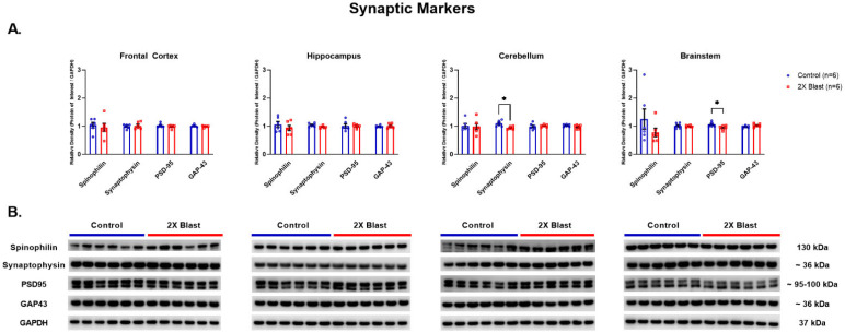
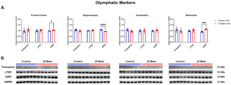
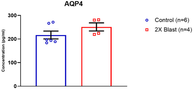
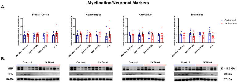
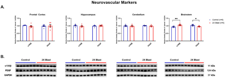
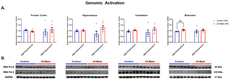
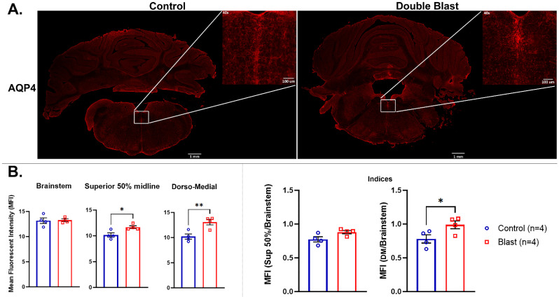
Explosive blasts are associated with neurological consequences as a result of blast waves impact on the brain. Yet, the neuropathologic and molecular consequences due to blast waves vs. blunt-TBI are not fully understood. An explosive-driven blast-generating system was used to reproduce blast wave exposure and examine pathological and molecular changes generated by primary wave effects of blast exposure. We assessed if pre- and post-synaptic (synaptophysin, PSD-95, spinophilin, GAP-43), neuronal (NF-L), glymphatic (LYVE1, podoplanin), myelin (MBP), neurovascular (AQP4, S100β, PDGF) and genomic (DNA polymerase-β, RNA polymerase II) markers could be altered across different brain regions of double blast vs. sham animals. Twelve male rats exposed to two consecutive blasts were compared to 12 control/sham rats. Western blot, ELISA, and immunofluorescence analyses were performed across the frontal cortex, hippocampus, cerebellum, and brainstem. The results showed altered levels of AQP4, S100β, DNA-polymerase-β, PDGF, synaptophysin and PSD-95 in double blast vs. sham animals in most of the examined regions. These data indicate that blast-generated changes are preferentially associated with neurovascular, glymphatic, and DNA repair markers, especially in the brainstem. Moreover, these changes were not accompanied by behavioral changes and corroborate the hypothesis for which an asymptomatic altered status is caused by repeated blast exposures.

Keywords: blast-wave primary effect; brain region-based blast-sensitivity; brainstem; molecular changes; neurodegeneration; subclinical status; traumatic brain injury.


Conflict of interest statement

The authors declare no conflict of interest.

Figures

Figure 1
Figure 1
Synaptic related protein expression changes. (A) Densitometric ratio of the levels of Spinophilin (single band at 130 kDa), Synaptophysin, PSD-95 (both visible bands evaluated together) and GAP-43 with respect to GAPDH as measured in FCtx, H, CRB and BS 15 days following an explosive-driven double blast exposure, n = 6 per group, * indicates p values < 0.05 as determined by 2-tailed, unpaired t-tests. Error bars represent standard error of the mean (SEM). (B) Representative western blots # for each antibody used. All gels were run in triplicate and data represents the average of 3 runs per sample. # For full length blots, see Supplementary Figures S1–S4.
Figure 2
Figure 2
Glymphatic related protein expression changes. (A) Densitometric ratio of the levels of Podoplanin, LYVE1, and AQP4 with respect to GAPDH as measured in the FCtx, H, CRB and BS 15 days following an explosive-driven double blast exposure, n = 6 per group, * indicates p values < 0.05, *** indicates p values < 0.001 and **** indicates p values < 0.0001, as determined by 2-tailed, unpaired t-tests. Error bars represent standard error of the mean (SEM). (B) Representative western blots # for each antibody used. All gels were run in triplicate and data represents the average of 3 runs per sample. # For full length blots, see Supplementary Figures S5–S7.
Figure 3
Figure 3
Plasma concentration of AQP4 following double blast exposure. Histogram representing the plasma concentration of AQP4 15 days following an explosive-driven double blast exposure, Ctl, n = 6, Blast, n = 4.
Figure 4
Figure 4
Myelin and neuronal related protein expression changes. (A) Densitometric ratio of the levels of MBP, isoforms 21.5 kDa and 18 kDa, (isoforms were evaluated together and separately) and NF-L with respect to GAPDH as measured in the FCtx, H, CRB and BS 15 days following an explosive-driven double blast exposure, n = 6 per group. Error bars represent standard error of the mean (SEM). (B) Representative western blots # for each antibody used. All gels were run in triplicate and data represents the average of 3 runs per sample. # For full length blots, see Supplementary Figures S8 and S9.
Figure 5
Figure 5
Neurovascular related protein expression changes. (A) Densitometric ratio of the levels of S100β and PDGF with respect to GAPDH as measured in the FCtx, H, CRB and BS 15 days following an explosive-driven double blast exposure, n = 6 per group, * indicates p values < 0.05 and ** indicates p values < 0.01 as determined by 2-tailed, unpaired t-tests. Error bars represent standard error of the mean (SEM). (B) Representative western blots # for each antibody used. All gels were run in triplicate and data represents the average of 3 runs per sample. # For full length blots, see Supplementary Figures S10 and S11.
Figure 6
Figure 6
Genomic activation/repair related protein expression changes. (A) Densitometric ratio of the levels of DNA Polymerase β and RNA Polymerase II with respect to GAPDH as measured in the FCtx, H, CRB and BS 15-days following an explosive-driven double blast exposure, n = 6 per group, ** indicates p values < 0.01 as determined by 2-tailed, unpaired t-tests. Error bars represent standard error of the mean (SEM). (B) Representative western blots # for each antibody used. All gels were run in triplicate and data represents the average of 3 runs per sample. # For full length blots, see Supplementary Figures S12 and S13.
Figure 7
Figure 7
AQP4 IF intensity changes in the BS following double blast exposure. AQP4 in control (Ctl.) versus double blast (2×B) rat BS. (A) AQP4-ir positive staining in the BS of a control rat not exposed to blast versus BS of a rat exposed to double blast, taken at 10× magnification. The insets at 40× magnification, defined as superior raphe obscurus-mfl, indicate the area (dorso-medial area) of MFI quantification. (B) Graphs showing MFI of total BS, 50% superior BS midline, and dorso-medial area including the superior portion of the raphe obscurus nuclei and mlf. * indicates p values < 0.05 and ** indicates p values < 0.01 as determined by 2-tailed, unpaired t-tests. Indices are MFI of each ROI over MFI of the total brainstem. IF = Immunofluorescence; BS = brainstem; mlf = medial longitudinal fasciculus; indices = IF intensity ratios between total BS/dorso-medial areas; MFI = mean fluorescent intensity.

References

    1. Hoge C., McGurk D., Thomas J., Cox A., Engel C., Castro C. Mild traumatic brain injury in U.S. soldiers returning from Iraq. N. Engl. J. Med. 2008;358:453–463. doi: 10.1056/NEJMoa072972. - DOI - PubMed
    1. Wallace D. Improvised explosive devices and traumatic brain injury: The military experience in Iraq and Afghanistan. Australas. Psychiatry. 2009;17:218–224. doi: 10.1080/10398560902878679. - DOI - PubMed
    1. Elder G., Mitsis E., Ahlers S., Cristian A. Blast-induced mild traumatic brain injury. Psychiatr. Clin. N. Am. 2010;33:757–781. doi: 10.1016/j.psc.2010.08.001. - DOI - PubMed
    1. Elder G.A., Stone J.R., Ahlers S.T. Effects of low-level blast exposure on the nervous system: Is there really a controversy? Front. Neurol. 2014;5:269. doi: 10.3389/fneur.2014.00269. - DOI - PMC - PubMed
    1. Nabity P.S., Jaramillo C.A., Resick P.A., McGeary C.A., Eapen B.C., Straud C.L., Hale W.J., Houle T.T., Litz B.T., Mintz J., et al. Consortium to Alleviate PTSD. Persistent posttraumatic headaches and functioning in veterans, Injury type can matter. Headache. 2021;61:1334–1341. doi: 10.1111/head.14210. - DOI - PubMed