Review
. 2023 Feb 11;13(2):258.
doi: 10.3390/bios13020258.

Recent Advances on Peptide-Based Biosensors and Electronic Noses for Foodborne Pathogen Detection

Affiliations
Review

Recent Advances on Peptide-Based Biosensors and Electronic Noses for Foodborne Pathogen Detection

Vanessa Escobar et al. Biosensors (Basel). .

Abstract

Foodborne pathogens present a serious issue around the world due to the remarkably high number of illnesses they cause every year. In an effort to narrow the gap between monitoring needs and currently implemented classical detection methodologies, the last decades have seen an increased development of highly accurate and reliable biosensors. Peptides as recognition biomolecules have been explored to develop biosensors that combine simple sample preparation and enhanced detection of bacterial pathogens in food. This review first focuses on the selection strategies for the design and screening of sensitive peptide bioreceptors, such as the isolation of natural antimicrobial peptides (AMPs) from living organisms, the screening of peptides by phage display and the use of in silico tools. Subsequently, an overview on the state-of-the-art techniques in the development of peptide-based biosensors for foodborne pathogen detection based on various transduction systems was given. Additionally, limitations in classical detection strategies have led to the development of innovative approaches for food monitoring, such as electronic noses, as promising alternatives. The use of peptide receptors in electronic noses is a growing field and the recent advances of such systems for foodborne pathogen detection are presented. All these biosensors and electronic noses are promising alternatives for the pathogen detection with high sensitivity, low cost and rapid response, and some of them are potential portable devices for on-site analyses.

Keywords: antimicrobial peptides; biosensors; electronic nose; foodborne pathogen; pathogenic detection; peptides; phage display.


Conflict of interest statement

The authors declare no conflict of interest.

Figures

Figure 1
Figure 1
Detection methodologies for foodborne pathogen detection in the literature in the period of 2002–2021. Values were obtained by searching “foodborne pathogen detection” and synonyms as keywords in Scopus, then classifying 2424 articles found in the literature by methodologies. Trends obtained by fitting a tendency curve and projecting it for the next 4 years.
Figure 2
Figure 2
Published articles on biosensors for the detection of foodborne pathogens in the literature in the period of 2002–2022. Values obtained by limiting “foodborne pathogen detection” and synonyms as a keywords article search in Scopus from 2002 to 2022 to biosensors, then classifying the 1167 articles by target foodborne pathogen and transduction techniques.
Figure 3
Figure 3
Different strategies for the design of foodborne pathogen-targeting peptides.
Figure 4
Figure 4
Schematic illustration of the potentiometric sandwich assay based on short antimicrobial peptide pairs for the detection of L. monocytogenes [118]. Copyright 2018, American Chemical Society.
Figure 5
Figure 5
(A) Schematic impedimetric set up of Magainin I immobilized on an interdigitated microelectrode array. (B) Magainin I in helical form, modified with a terminal cysteine residue and with clearly defined hydrophobic and hydrophilic faces. (C) Detection of bacteria achieved via binding of target cells to the immobilized AMPs. (D) Optical image of the interdigitated microelectrode array (scale bar: 50 μm) [120]. Copyright 2010, National Academy of Sciences.
Figure 6
Figure 6
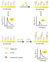
Schematic representation of the EIS biosensor based on magnetic nanoparticles functionalized with antimicrobial peptides. (A) Functionalization of MNPs with Melittin. (B) Capture of bacteria by MNPs-MLT, and magnetic separation of bacteria from the sample matrix. (C) EIS detection [121]. Copyright 2019, Elsevier.
Figure 7
Figure 7
Schematic setup for norovirus detection using a peptide-based EIS biosensor. (A) The novel peptides were immobilized by the formation of SAMs on the Au working electrode. (B) Using a working buffer solution, the dropped norovirus on the Au working electrode was then measured, along with its strength affinity, by using an EIS analysis [40]. Copyright 2019, Elsevier.
Figure 8
Figure 8
Schematic fabrication process of the PEC sensing platform for the detection of E. coli O157:H7. (I) Assembly of UCNPs@SiO2@Ag/C-g-C3N4, Magainin I and BSA onto PWE (II) PEC response of fabricated sensing platform under NIR light of 980 nm (a) photograph of modified PWE, Ag/AgCl reference electrode and carbon counter electrode assembled randomly on paper, (b) lab-on-paper PEC platform [122]. Copyright 2022, Elsevier.
Figure 9
Figure 9
Schematic diagram of the simultaneous detection of E. coli O157:H7, L. monocytogenes and B. melitensis 16M based on phage-displayed peptides and the fluorescence spectroscopy set up [84]. Copyright 2020, Elsevier.
Figure 10
Figure 10
Schematic illustration of the isolation of the pVIII-coated protein fused with an S. aureus-specific octapeptide and the formation of CS-AuNPs@fusion-pVIII [86]. Copyright 2016, Elsevier.
Figure 11
Figure 11
Schematic illustration of the AMP-based colorimetric bioassay for the detection of E. coli O157:H7 [130]. Copyright 2017, Royal Society of Chemistry.
Figure 12
Figure 12
Schematic illustration of colorimetric detection of V. parahaemolyticus. (A) V. parahaemolyticus-specific pVIII fusion protected by tert-Butyl carbamate. (B) The C-terminal end of pVIII fusion was activated by EDC/NHS, bioconjugated with MnO2 NSs, and the N-terminal of pVIII fusion was deprotected. (C) MnO2NS@ pVIII fusion as sandwich immunoassay tags for V. parahaemolyticus detection [85]. Copyright 2018, Royal Society of Chemistry.
Figure 13
Figure 13
Schematic SPRI set up and principal component analysis discrimination between S. aureus, S. typhimurium, S. epidermis, E. coli and L. monocytogenes [111]. Copyright 2019, Elsevier.
Figure 14
Figure 14
Schematic illustration of optical fiber SPR detection of E. coli O157:H7 [114]. Copyright 2018, Elsevier.
Figure 15
Figure 15
Schematic electrochemiluminescence biosensor fabrication and ECL determination of E. coli O157:H7 [133]. Copyright 2015, Elsevier.
Figure 16
Figure 16
The schematic illustration of the nanomechanical peptide-based biosensors and its multi-mode of operation (A) BMC filled with bacteria supported on a silicon substrate. The BMC was coated with a bacteria-targeted receptor and irradiated with a specific wavelength of tunable infrared light. (B) Scanning electron microscopy (SEM) image of the cross-section of an inlet, through which an aqueous solution of bacteria is loaded. (C) Cross-section of the 32 μm wide microchannel of the cantilever functionalized with either a mAb or Leucocin A, which acted specifically against L. monocytogenes. (D) Fluorescent image from the top side of the BMC, filled with bacteria. (E) SEM image of the tip of the BMC. (F) When the bacteria inside the BMC absorbs infrared light, local heat is generated that results in the nanomechanical deflection of the BMC. (G) The resonance frequency is sensitive to the increased mass caused by the adsorption of bacteria inside the BMC. (H) When the BMC is illuminated with a certain range of infrared light, a plot of the nanomechanical deflection of the BMC shows the wavelength where the bacteria absorb infrared light [135]. Copyright 2016, Springer Nature.
Figure 17
Figure 17
The electronic nose for the detection of TMA. (A) ORPs were self-assembled on the surface of SWNTs during the treatment of ORP-suspended deionized water solutions. The ORPs were immobilized by π–π stacking of aromatic rings at their C-terminus and attracted TMA molecules very near to the SWNTs. (B) Atomic force microscopy images of bare and ORP-immobilized SWNT channels on the fabricated sensor devices. (C) Height profiles of specific cross sections (white dashed lines in (B)). The height of SWNTs increased by 2–3 nm after immobilization of the ORPs. [162]. Copyright 2013, Elsevier.
Figure 18
Figure 18
Schematic setup of CNT-FET for the detection of Salmonella contamination in ham and real-time detection of Salmonella contamination in sliced ham [165]. Copyright 2016, American Chemical Society.

References

    1. Abebe E., Gugsa G., Ahmed M. Review on Major Food-Borne Zoonotic Bacterial Pathogens. J. Trop. Med. 2020;2020:4674235. doi: 10.1155/2020/4674235. - DOI - PMC - PubMed
    1. World Health Organization (WHO) Estimates of the Global Burden of Foodborne Diseases: Foodborne Disease Burden Epidemiology Reference Group 2007–2015. World Health Organization; Geneva, Switzerland: 2015.
    1. Yadav S., Kapley A. Antibiotic Resistance: Global Health Crisis and Metagenomics. Biotechnol. Rep. 2021;29:e00604. doi: 10.1016/j.btre.2021.e00604. - DOI - PMC - PubMed
    1. Desta Sisay M.A. A Review on Major Food Borne Bacterial Illnesses. J. Trop. Dis. 2015;3:176. doi: 10.4172/2329-891X.1000176. - DOI
    1. European Food Safety Authority. European Centre for Disease Prevention and Control The European Union One Health 2020 Zoonoses Report. EFSA J. 2021;19:e06971. doi: 10.2903/j.efsa.2021.6971. - DOI - PMC - PubMed

LinkOut - more resources