Review
. 2023 Feb 9;13(4):645.
doi: 10.3390/diagnostics13040645.

Photon-Counting Computed Tomography (PCCT): Technical Background and Cardio-Vascular Applications

Affiliations
Review

Photon-Counting Computed Tomography (PCCT): Technical Background and Cardio-Vascular Applications

Antonella Meloni et al. Diagnostics (Basel). .

Abstract

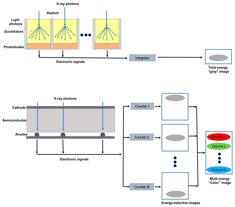
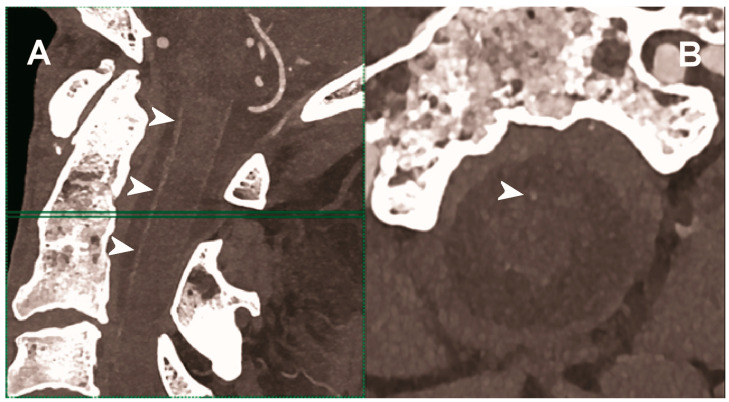
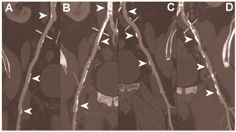
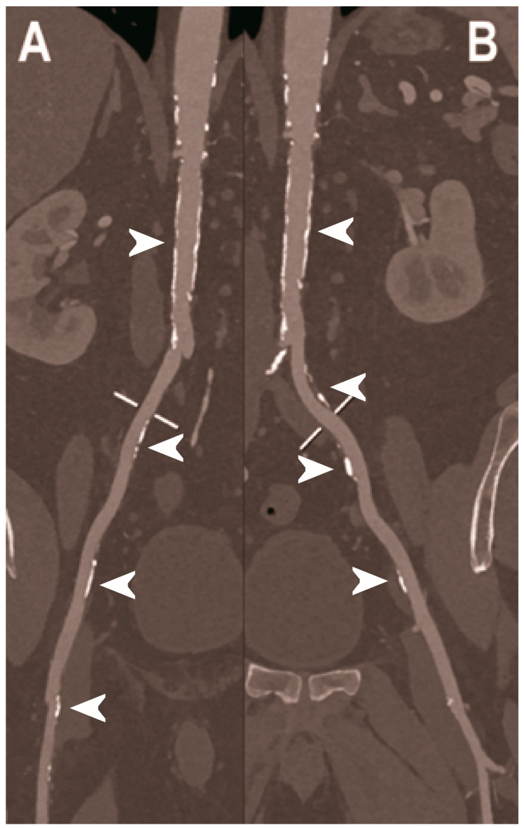
Photon-counting computed tomography (PCCT) is a new advanced imaging technique that is going to transform the standard clinical use of computed tomography (CT) imaging. Photon-counting detectors resolve the number of photons and the incident X-ray energy spectrum into multiple energy bins. Compared with conventional CT technology, PCCT offers the advantages of improved spatial and contrast resolution, reduction of image noise and artifacts, reduced radiation exposure, and multi-energy/multi-parametric imaging based on the atomic properties of tissues, with the consequent possibility to use different contrast agents and improve quantitative imaging. This narrative review first briefly describes the technical principles and the benefits of photon-counting CT and then provides a synthetic outline of the current literature on its use for vascular imaging.

Keywords: CT angiography; cardiac CT; photon-counting CT; photon-counting detectors.


Conflict of interest statement

The authors declare no conflict of interest.

Figures

Figure 1
Figure 1
Schematic representation of an energy integrating detector (top) and of a photon-counting detector directly converting X-rays into an electrical signal (bottom). The photon-counting design allows the generation of energy-selective images, from which a set of material concentration maps can be obtained. Material concentration maps can then be combined in different ways to obtain monochromatic images, virtual non-contrast images, or material-specific color-overlay images.
Figure 2
Figure 2
Cardiac CT using photon-counting computed tomography. The figure shows advanced multiplanar reconstructions of a coronary tree derived from a Photon-Counting CT (Scanner: NAEOTOM Alpha, Siemens) acquisition (AC). In (A) we can see the right coronary artery with 2 stents (arrowheads), one proximal and one distal (much smaller), without any issue in the intrastent visualization of the arterial lumen. In (B), the left anterior descending coronary artery is depicted along its entire course (down to and beyond the left ventricular apex) with great detail and an evident deep intramyocardial course in the middle segment of the vessel (arrowhead). In (C), the left circumflex coronary artery also shows a stent with perfect intrastent visualization and patency (arrowhead).
Figure 3
Figure 3
Carotid CT angiography using photon-counting computed tomography. The figure shows advanced reconstructions of a carotid artery tree derived from a photon-counting CT (Scanner: NAEOTOM Alpha, Siemens) acquisition (AD). In (A), a longitudinal multiplanar reconstruction shows the right common and internal carotid artery with no significant luminal stenosis and a predominantly calcified atherosclerotic plaque at the carotid bifurcation (arrowhead). In (B), a longitudinal multiplanar reconstruction shows the left common and internal carotid artery with a significant luminal stenosis in the post-bulbar region of the internal carotid artery (arrowhead). In (C) the quantitative assessment of the stenosis (arrowhead) and in (D) the 3-dimensional volume rendering of the lesion (arrowhead).
Figure 4
Figure 4
Carotid CT angiography using photon-counting computed tomography. The figure shows advanced reconstructions of a carotid artery tree derived from a photon-counting CT (Scanner: NAEOTOM Alpha, Siemens) acquisition (AD). In (A,B), a longitudinal multiplanar reconstruction shows the right common and internal carotid artery with no significant luminal stenosis and a mild predominantly calcified atherosclerotic plaque at the internal carotid origin (arrowhead). In (C,D), a longitudinal multiplanar reconstruction shows the left common and internal carotid artery with a minimal arterial wall irregularity at the internal carotid origin (arrowhead). What is a bit unusual is to be able to see the thickness of the arterial wall at this level in a case with very mild atherosclerotic disease.
Figure 5
Figure 5
Carotid CT angiography using photon-counting computed tomography. The figure shows advanced MIP reconstructions of a intracervical artery tree derived from a photo-counting CT (Scanner: NAEOTOM Alpha, Siemens) acquisition. In (A) a sagittal median view of the cervical region showing the course of the anterior spinal artery (normally not visible) in the ventral portion of the rachidial channel (arrowheads). In (B) the axial image at the level of the green plane showed on the left panel with the axial view of the anterior spinal artery (arrowhead).
Figure 6
Figure 6
Abdominal CT angiography using photon-counting computed tomography. The figure shows advanced multiplanar reconstructions without and with MIP algorithm of a distal abdominal aorta and ilio-femoral arterial axes derived from a photon-counting CT (Scanner: NAEOTOM Alpha, Siemens) acquisition (A,B right; C,D left). The projection start in the abdominal aorta carrefour and end in the right/left common femoral artery. There are massive calcifications along the common iliac arteries; however, both MPRs (A,C) and MIPs (B,D) are so sharply defining the edges of the structures that lumen assessment is not compromised (arrowheads).
Figure 7
Figure 7
Abdominal CT angiography using photon-counting computed tomography. The figure shows advanced multiplanar reconstructions of an abdominal aorta and ilio-femoral arterial axes derived from a photon-counting CT (Scanner: NAEOTOM Alpha, Siemens) acquisition (A,B). In (A) the projection starts in the abdominal aorta at the level of thoraco-abdominal junction and ends in the right common femoral artery while in (B) it ends in the left common femoral artery. While there are significant calcifications along the vessels (arrowheads in A and B), the vessel wall is so sharp that the fact that there is no lumen reduction appears to be quite natural as compared to the common blooming effect seen with conventional energy-integrating detectors.

References

    1. Libby P., Ridker P.M. Inflammation and atherosclerosis: Role of C-reactive protein in risk assessment. Am. J. Med. 2004;116:9–16. doi: 10.1016/j.amjmed.2004.02.006. - DOI - PubMed
    1. Aziz M., Yadav K. Pathogenesis of atherosclerosis a review. Med. Clin. Rev. 2016;2:1–6.
    1. Virani S.S., Alonso A., Aparicio H.J., Benjamin E.J., Bittencourt M.S., Callaway C.W., Carson A.P., Chamberlain A.M., Cheng S., Delling F.N., et al. Heart Disease and Stroke Statistics-2021 Update: A Report From the American Heart Association. Circulation. 2021;143:e254–e743. doi: 10.1161/CIR.0000000000000950. - DOI - PubMed
    1. Andreini D., Martuscelli E., Guaricci A.I., Carrabba N., Magnoni M., Tedeschi C., Pelliccia A., Pontone G. Clinical recommendations on Cardiac-CT in 2015: A position paper of the Working Group on Cardiac-CT and Nuclear Cardiology of the Italian Society of Cardiology. J. Cardiovasc. Med. 2016;17:73–84. doi: 10.2459/JCM.0000000000000318. - DOI - PubMed
    1. Heseltine T.D., Murray S.W., Ruzsics B., Fisher M. Latest Advances in Cardiac CT. Eur. Cardiol. 2020;15:1–7. doi: 10.15420/ecr.2019.14.2. - DOI - PMC - PubMed

LinkOut - more resources