Review
. 2023 Feb 11;13(4):677.
doi: 10.3390/diagnostics13040677.

Towards OCT-Guided Endoscopic Laser Surgery-A Review

Affiliations
Review

Towards OCT-Guided Endoscopic Laser Surgery-A Review

Ajay Gunalan et al. Diagnostics (Basel). .

Abstract

Optical Coherence Tomography (OCT) is an optical imaging technology occupying a unique position in the resolution vs. imaging depth spectrum. It is already well established in the field of ophthalmology, and its application in other fields of medicine is growing. This is motivated by the fact that OCT is a real-time sensing technology with high sensitivity to precancerous lesions in epithelial tissues, which can be exploited to provide valuable information to clinicians. In the prospective case of OCT-guided endoscopic laser surgery, these real-time data will be used to assist surgeons in challenging endoscopic procedures in which high-power lasers are used to eradicate diseases. The combination of OCT and laser is expected to enhance the detection of tumors, the identification of tumor margins, and ensure total disease eradication while avoiding damage to healthy tissue and critical anatomical structures. Therefore, OCT-guided endoscopic laser surgery is an important nascent research area. This paper aims to contribute to this field with a comprehensive review of state-of-the-art technologies that may be exploited as the building blocks for achieving such a system. The paper begins with a review of the principles and technical details of endoscopic OCT, highlighting challenges and proposed solutions. Then, once the state of the art of the base imaging technology is outlined, the new OCT-guided endoscopic laser surgery frontier is reviewed. Finally, the paper concludes with a discussion on the constraints, benefits and open challenges associated with this new type of surgical technology.

Keywords: OCT; endoscopy; laser surgery; theranostics systems.


Conflict of interest statement

The authors declare no conflict of interest.

Figures

Figure 1
Figure 1
Comparison between Spectral Domain OCT (SD-OCT) and Swept Source OCT (SS-OCT) by [9]. Figure licensed under CC BY 4.0.
Figure 2
Figure 2
Scanning geometries of endoscopic OCT systems: side, circumferential and forward scanning.
Figure 3
Figure 3
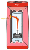
Schematic of a 3D-printed OCT endoscope inside an artery by [18]. Figure licensed under CC BY 4.0.
Figure 4
Figure 4
Example of a micromotor-based balloon catheter for OCT imaging, used with permission from [28] © The Optical Society.
Figure 5
Figure 5
Schematic diagram of an OCT imaging system for pediatric laryngoscopy with a proximal scanning mechanism by [32]. Figure licensed under CC BY 4.0.
Figure 6
Figure 6
Examples of distal scanning endoscopic OCT probes based on a mirror and on a fiber scanner.
Figure 7
Figure 7
Proximal scanning system for an endoscopic OCT. Light from a stationary source fiber is coupled to a rotating endoscopic probe, used with permission from [12] © The Optical Society.
Figure 8
Figure 8
OCT-guided laser surgery systems based on double-clad fibers. (a) Different types of optical fibers: single-mode fiber (SMF), double-clad fiber (DCF) and multi-mode fiber (MMF). (b) Experimental setup proposed for co-localized OCT imaging and laser therapy, used with permission from [38] © The Optical Society.
Figure 9
Figure 9
OCT-guided laser surgery systems based on dichroic mirrors. (a) Control loop scheme of the OCT-guided laser cochleostomy by [46] is licensed under CC BY 3.0. (b) Data flow and representation in the smart laser surgical system, used with permission from [40] © The Wiley.
Figure 10
Figure 10
Combined miniature B-scan OCT and surgical laser in an intraocular probe, used with permission from [52] © The Wiley. (A) Picture of the device. (B) Magnified picture showing the support strucutre (white arrow) and the combined OCT (red arrow) and laser (blue arrow) probe tip. (C) Details of the optical components that allow co-planar ablation and imaging. (D) Diagram of the combined OCT and laser probe tip showing the scanning optical fiber.

References

    1. Siegel R., Naishadham D., Jemal A. Cancer statistics, 2012. CA A Cancer J. Clin. 2012;62:10–29. doi: 10.3322/caac.20138. - DOI - PubMed
    1. Qiu Z., Piyawattanamatha W. New Endoscopic Imaging Technology Based on MEMS Sensors and Actuators. Micromachines. 2017;8:210. doi: 10.3390/mi8070210. - DOI - PMC - PubMed
    1. Sun J., Xie H. MEMS-Based Endoscopic Optical Coherence Tomography. Int. J. Opt. 2011;2011:825629. doi: 10.1155/2011/825629. - DOI
    1. Rollins A.M., Izatt J.A. Optimal interferometer designs for optical coherence tomography. Opt. Lett. 1999;24:1484–1486. doi: 10.1364/OL.24.001484. - DOI - PubMed
    1. Aumann S., Donner S., Fischer J., Müller F. Optical Coherence Tomography (OCT): Principle and Technical Realization. In: Bille J.F., editor. High Resolution Imaging in Microscopy and Ophthalmology: New Frontiers in Biomedical Optics. Springer International Publishing; Cham, Switzerland: 2019. pp. 59–85. - DOI - PubMed

LinkOut - more resources