. 2023 Feb 13;13(4):711.
doi: 10.3390/diagnostics13040711.

Low-Dose CT Fluoroscopy-Guided Drainage of Deep Pelvic Fluid Collections after Colorectal Cancer Surgery: Technical Success, Clinical Outcome and Safety in 40 Patients

Affiliations

Low-Dose CT Fluoroscopy-Guided Drainage of Deep Pelvic Fluid Collections after Colorectal Cancer Surgery: Technical Success, Clinical Outcome and Safety in 40 Patients

Robert Stahl et al. Diagnostics (Basel). .

Abstract

Purpose: To assess the technical (TS) and clinical success (CS) of CT fluoroscopy-guided drainage (CTD) in patients with symptomatic deep pelvic fluid collections following colorectal surgery.

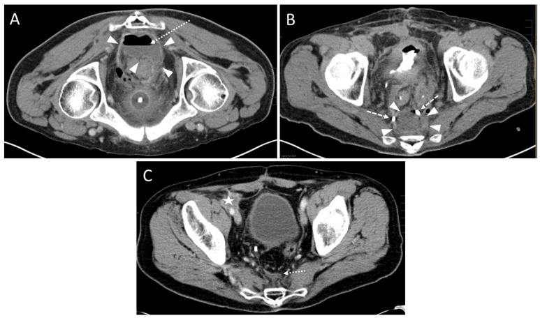
Methods: A retrospective analysis (years 2005 to 2020) comprised 43 drain placements in 40 patients undergoing low-dose (10-20 mA tube current) quick-check CTD using a percutaneous transgluteal (n = 39) or transperineal (n = 1) access. TS was defined as sufficient drainage of the fluid collection by ≥50% and the absence of complications according to the Cardiovascular and Interventional Radiological Society of Europe (CIRSE). CS comprised the marked reduction of elevated laboratory inflammation parameters by ≥50% under minimally invasive combination therapy (i.v. broad-spectrum antibiotics, drainage) within 30 days after intervention and no surgical revision related to the intervention required.

Results: TS was gained in 93.0%. CS was obtained in 83.3% for C-reactive Protein and in 78.6% for Leukocytes. In five patients (12.5%), a reoperation due to an unfavorable clinical outcome was necessary. Total dose length product (DLP) tended to be lower in the second half of the observation period (median: years 2013 to 2020: 544.0 mGy*cm vs. years 2005 to 2012: 735.5 mGy*cm) and was significantly lower for the CT fluoroscopy part (median: years 2013 to 2020: 47.0 mGy*cm vs. years 2005 to 2012: 85.0 mGy*cm).

Conclusions: Given a minor proportion of patients requiring surgical revision due to anastomotic leakage, the CTD of deep pelvic fluid collections is safe and provides an excellent technical and clinical outcome. The reduction of radiation exposition over time can be achieved by both the ongoing development of CT technology and the increased level of interventional radiology (IR) expertise.

Keywords: CT-guided drainage; clinical outcome; colorectal surgery; pelvic fluid collection; technical outcome.


Conflict of interest statement

The authors declare that they have no conflicts of interest.

Figures

Figure 1
Figure 1
Flow chart of the patient selection process.
Figure 2
Figure 2
Three cases of pelvic fluid collections after colorectal cancer surgery. Three different access trajectories are depicted: Upper row: Transpiriform access path. of a 69 year-old male with a history of rectal carcinoma (T2 tumor stage) after deep anterior rectal resection including total mesorectal excision and the creation of a loop-iliostomy. Partial anastomotic insufficiency with concomitant presacral abscess was observed after two months and initially treated using endoluminal vacuum therapy. Subsequently, a CT fluoroscopy guided drain was inserted after four months (Leukocytes 8.3 × 10⁹/L; CRP: 1.6 mg/dL). (A) A preinterventional contrast enhanced CT shows a presacral fluid collection with marked rim enhancement (arrowheads). Loculated small gas collection indicating a fistula and superinfection. (B) CT fluoroscopy-guided insertion of a 10 F drain (arrow). Arrowheads: fluid collection; dotted arrow: loculated gas collection. (C) Postinterventional control scan. Reduced size of the fluid collection (arrowheads) after aspiration. Arrow: drain; dashed arrow: loop-formation of the distal drain segment. Microbiological analysis revealed infection with Enterococcus faecalis, Haemophilus parainfluenzae, Escherichia coli, Proteus vulgaris, Citrobacter koseri, and Bacteroides uniformis. Middle row: Infrapiriform access path. A 51 year-old male with history of hepatic metastatic rectal cancer (T3 tumor stage) and deep anterior rectal resection with Hartmann’s operation after neoadjuvant chemotherapy with FOLFOX4/FOLFIRI schema. Two months after surgery the patient presented with elevated temperature and fever spikes up to 39.5 degrees C (Leukocytes 17.4 × 10⁹/L; CRP: 5.4 mg/dL). A CT fluoroscopy-guided drain was applied. (D) A preinterventional contrast enhanced CT showed a presacral fluid collection with marked rim enhancement (arrowheads). (E) CT fluoroscopy placement of an 8 F drain (arrow) in semi-prone position. Arrowheads: fluid collection. (F) Postoperative control scan. After aspiration of the abscess cavity, a loculated small collection of air could be seen (dotted arrow). The size of the fluid collection (arrowheads) was reduced. A microbiological analysis revealed an infection with Bacteroides fragilis. Lower row: Transperineal access path. of a 74 year-old male with history of a deep-seated rectal carcinoma (yT3 tumor stage) after laparoscopic abdominoperineal rectum resection. The patient had fever of up to 39 degrees with recurrent spikes since the 9th postoperative day. Fifteen days after surgery, a CT revealed postoperative fluid retention in the pelvic floor extending to the penile root (Leukocytes 6.8 × 10⁹/L; CRP: 6.9 mg/dL). (G) A contrast enhanced planning CT scan in the prone position did not show rim enhancement or air bubbles within the collection (arrowheads). Asterisk: penile root. (H) CT fluoroscopy-guided insertion of an 8 F single lumen pigtail drain (arrow) into the perineal region using the Trocar-technique was performed. Arrowheads: fluid collection; asterisk: penile root. (I) Complete resolution of the fluid collection after aspiration was seen in the control scan. Arrow: drain; dashed arrow: loop-formation of the distal drain segment; asterisk: penile root. A microbiological analysis revealed infection with Proteus mirabilis.
Figure 3
Figure 3
A 59-year-old male with history of rectal cancer (T3 tumor stage) after deep anterior rectal resection including total mesorectal excision and creation of a loop-iliostomy. A control CT scan after 9 weeks revealed a presacral contrast enhancing fluid collection. The patient had no clinical symptoms (Leukocytes: 4.1 × 10⁹/L; CRP: 0.6 mg/dL). (A) Preinterventional contrast enhanced CT scan in prone position showing the presacral fluid collection with rim enhancement (arrowheads). (B) Insertion of an 8 F drain (arrow) into the abscess formation (arrowheads). Only 20 mL of fluid could be aspirated. A microbiological analysis revealed an encapsulated hematoma infected with peptostreptococcus species. (C) Six weeks after the intervention, the fluid accumulation (arrowheads) showed a slight regression in size. Hypodense scar tissue (dotted arrows) had developed. (D) After one year the hematoma was completely resolved. Dotted arrows: scar tissue.
Figure 4
Figure 4
A 67-year-old male with a history of rectal carcinoma (T4 tumor stage) after deep anterior rectal resection including total mesorectal excision and the creation of a loop-iliostomy. In addition, a ureter reimplantation due to tumor infiltration in the bladder and an iliac lymph node dissection was performed. Five days after the operation a small anastomotic insufficiency was diagnosed and treated by flushing. Three weeks later a CT fluoroscopy-guided drain placement was performed due to recurrent fever spikes up to 38.5 degrees. (A) A preinterventional scan showed a presacral fluid collection (arrowheads) with air/fluid level (dotted arrow). (B) After the CT fluoroscopy-guided insertion of an 8 F drain, purulent fluid could be partially aspirated and a postinterventional control scan showed a moderate reduction of the abscess formation (arrowheads). Dashed arrow: loop-formation of the distal drain segment. (C) After one year, the complete resolution of presacral abscess formation was observed. Dotted arrow: small scar tissue. However, a newly appeared soft tissue mass in terms of a peritoneal tumor relapse occurred in the right parailiac region (asterisk).
Figure 5
Figure 5
Development of laboratory parameters within 30 days after the intervention in subjects (n = 19) with no evidence of further surgical interventions or complications in the patient record.

Similar articles

Cited by

References

    1. Baidoun F., Elshiwy K., Elkeraie Y., Merjaneh Z., Khoudari G., Sarmini M.T., Gad M., Al-Husseini M., Saad A. Colorectal Cancer Epidemiology: Recent Trends and Impact on Outcomes. Curr. Drug Targets. 2021;22:998–1009. doi: 10.2174/1389450121999201117115717. - DOI - PubMed
    1. Brenner H., Kloor M., Pox C.P. Colorectal cancer. Lancet. 2014;383:1490–1502. doi: 10.1016/S0140-6736(13)61649-9. - DOI - PubMed
    1. Sinicrope F.A. Increasing Incidence of Early-Onset Colorectal Cancer. N. Engl. J. Med. 2022;386:1547–1558. doi: 10.1056/NEJMra2200869. - DOI - PubMed
    1. Mármol I., Sánchez-de-Diego C., Pradilla Dieste A., Cerrada E., Rodriguez Yoldi M.J. Colorectal Carcinoma: A General Overview and Future Perspectives in Colorectal Cancer. Int. J. Mol. Sci. 2017;18:197. doi: 10.3390/ijms18010197. - DOI - PMC - PubMed
    1. Haraldsdottir S., Einarsdottir H.M., Smaradottir A., Gunnlaugsson A., Halfdanarson T.R. Colorectal cancer—Review. Laeknabladid. 2014;100:75–82. doi: 10.17992/lbl.2014.02.531. - DOI - PubMed