. 2023 Feb 16;13(4):751.
doi: 10.3390/diagnostics13040751.

Left Bronchial Isomerism with Right-Sided Tracheal Bronchus: A Rare Case Report

Affiliations

Left Bronchial Isomerism with Right-Sided Tracheal Bronchus: A Rare Case Report

Kyungsoo Bae et al. Diagnostics (Basel). .

Abstract

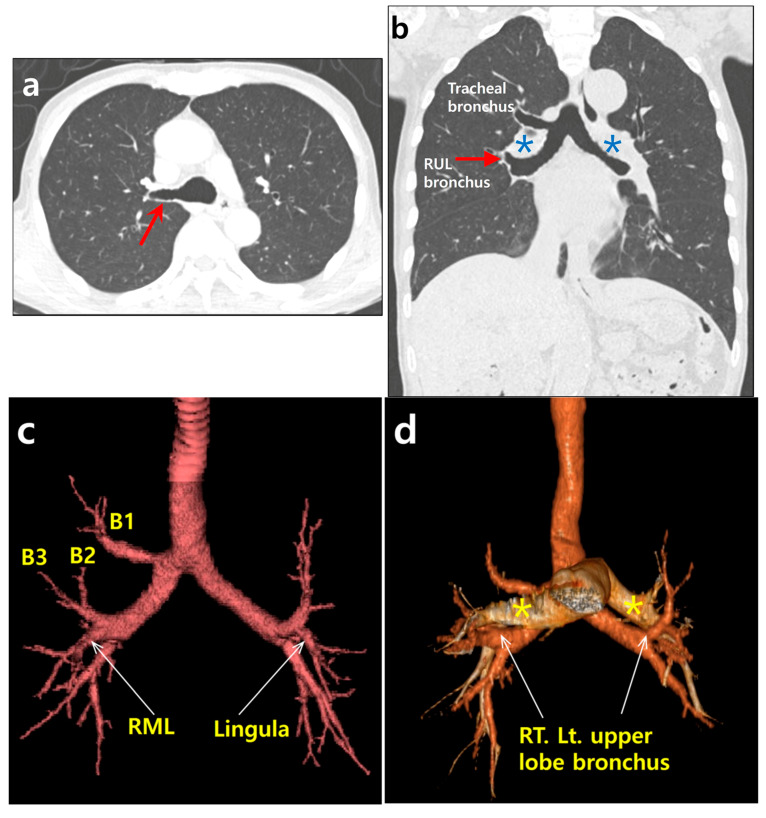
The tracheal bronchus is a congenital bronchial branching anomaly defined as an aberrant bronchus arising in either the trachea or a main bronchus. Left bronchial isomerism is characterized by two bilobed lungs, bilateral long main bronchi, and both pulmonary arteries passing superiorly to their respective upper lobe bronchi. Left bronchial isomerism with a right-sided tracheal bronchus is a very rare combination of tracheobronchial anomalies. It has not been previously reported. We present multi-detector CT findings of a left bronchial isomerism with a right-sided tracheal bronchus of a 74 year old man.

Keywords: bronchi; bronchial isomerism; computed tomography; trachea; tracheal bronchus.


Conflict of interest statement

The authors declare no conflict of interest.

Figures

Figure 1
Figure 1
A 74 year old man with tracheal bronchus and left bronchial isomerism. (a) An axial chest CT image with lung window setting showing an aberrant bronchus (tracheal bronchus, arrow) arising from the right main bronchus at the level of carina. (b) A coronal, reformatted chest CT image with lung window setting showing symmetrical long both main bronchi resembling the left bronchus morphologically. Right upper lobe bronchus (RUL) is located below the point where the pulmonary artery (asterisk) crosses the main bronchus as on the left (hyparterial). Note tracheal bronchus directing towards the right upper lobe. (c) Reconstructed, 3D CT image of the tracheobronchial tree revealing both upper lobe bronchi in the same position. Similar to lingular division (arrow) of the left upper lobe bronchus, the right middle lobe bronchus (RML, arrow) arises with the right upper lobe bronchus. Displaced apical segmental bronchus (B1, thin arrow) arises from tracheal bronchus. Posterior and anterior segmental bronchi (B2 and B3) arise from right upper lobe bronchus. (d) Superimposed 3D CT image of the tracheobronchial tree and pulmonary arteries (asterisks) showing that both upper lobe bronchi (arrows) are hyparterial. (e) Sagittal reformatted CT images with lung window setting showing bilobed left and right lungs (arrows indicate both major fissures). There is no minor fissure in the right lung.
Figure 1
Figure 1
A 74 year old man with tracheal bronchus and left bronchial isomerism. (a) An axial chest CT image with lung window setting showing an aberrant bronchus (tracheal bronchus, arrow) arising from the right main bronchus at the level of carina. (b) A coronal, reformatted chest CT image with lung window setting showing symmetrical long both main bronchi resembling the left bronchus morphologically. Right upper lobe bronchus (RUL) is located below the point where the pulmonary artery (asterisk) crosses the main bronchus as on the left (hyparterial). Note tracheal bronchus directing towards the right upper lobe. (c) Reconstructed, 3D CT image of the tracheobronchial tree revealing both upper lobe bronchi in the same position. Similar to lingular division (arrow) of the left upper lobe bronchus, the right middle lobe bronchus (RML, arrow) arises with the right upper lobe bronchus. Displaced apical segmental bronchus (B1, thin arrow) arises from tracheal bronchus. Posterior and anterior segmental bronchi (B2 and B3) arise from right upper lobe bronchus. (d) Superimposed 3D CT image of the tracheobronchial tree and pulmonary arteries (asterisks) showing that both upper lobe bronchi (arrows) are hyparterial. (e) Sagittal reformatted CT images with lung window setting showing bilobed left and right lungs (arrows indicate both major fissures). There is no minor fissure in the right lung.

Similar articles

Cited by

References

    1. Wong L.M., Cheruiyot I., de Oliveira M.H.S., Keet K., Tomaszewski K.A., Walocha J.A., Tubbs S., Henry B.M. Congenital Anomalies of the Tracheobronchial Tree: A Meta-Analysis and Clinical Considerations. Ann. Thorac. Surg. 2021;112:315–325. doi: 10.1016/j.athoracsur.2020.08.060. - DOI - PubMed
    1. Ghaye B., Szapiro D., Fanchamps J.M., Dondelinger R.F. Congenital bronchial abnormalities revisited. Radiographics. 2001;21:105–119. doi: 10.1148/radiographics.21.1.g01ja06105. - DOI - PubMed
    1. Wooten C., Patel S., Cassidy L., Watanabe K., Matusz P., Tubbs R.S., Loukas M. Variations of the tracheobronchial tree: Anatomical and clinical significance. Clin. Anat. 2014;27:1223–1233. doi: 10.1002/ca.22351. - DOI - PubMed
    1. Applegate K.E., Goske M.J., Pierce G., Murphy D. Situs revisited: Imaging of the heterotaxy syndrome. Radiographics. 1999;19:837–852; discussion 53–54. doi: 10.1148/radiographics.19.4.g99jl31837. - DOI - PubMed
    1. Pandey N.N., Verma M., Jagia P., Gupta S.K. Bilateral tracheal bronchi in a patient with right isomerism. J. Cardiovasc. Comput. Tomogr. 2022;16:e23–e24. doi: 10.1016/j.jcct.2021.11.010. - DOI - PubMed

LinkOut - more resources