. 2023 Feb 17;13(4):769.
doi: 10.3390/diagnostics13040769.

Lung Dual-Energy CT Perfusion Blood Volume as a Marker of Severity in Chronic Thromboembolic Pulmonary Hypertension

Affiliations

Lung Dual-Energy CT Perfusion Blood Volume as a Marker of Severity in Chronic Thromboembolic Pulmonary Hypertension

Salim A Si-Mohamed et al. Diagnostics (Basel). .

Abstract

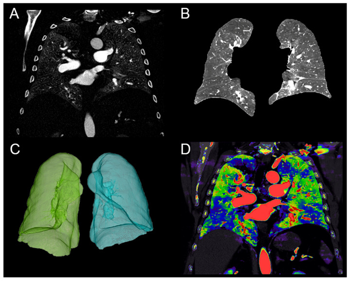
In chronic thromboembolic pulmonary hypertension (CTEPH), assessment of severity requires right heart catheterization (RHC) through cardiac index (CI). Previous studies have shown that dual-energy CT allows a quantitative assessment of the lung perfusion blood volume (PBV). Therefore, the objective was to evaluate the quantitative PBV as a marker of severity in CTEPH. In the present study, thirty-three patients with CTEPH (22 women, 68.2 ± 14.8 years) were included from May 2017 to September 2021. Mean quantitative PBV was 7.6% ± 3.1 and correlated with CI (r = 0.519, p = 0.002). Mean qualitative PBV was 41.1 ± 13.4 and did not correlate with CI. Quantitative PBV AUC values were 0.795 (95% CI: 0.637-0.953, p = 0.013) for a CI ≥ 2 L/min/m2 and 0.752 (95% CI: 0.575-0.929, p = 0.020) for a CI ≥ 2.5 L/min/m2. In conclusion, quantitative lung PBV outperformed qualitative PBV for its correlation with the cardiac index and may be used as a non-invasive marker of severity in CTPEH patients.

Keywords: X-ray computed/methods; comparative study; lung; perfusion; tomography.


Conflict of interest statement

The authors declare no conflict of interest.

Figures

Figure 1
Figure 1
Quantification of the lung perfusion blood volume (PBV) in a 64-year-old male patient with chronic thromboembolic pulmonary hypertension (CTEPH). (A) Coronal image of the lung iodine density map. (B) Coronal image after lung segmentation with the semi-automatic software COPD (IntelliSpace Portal; Philips Healthcare®) allowing the exclusion of the main pulmonary vessels. (C) Volumetric segmentation of the lungs. (D) Overlay coronal image of the conventional and iodine density maps representing the defect perfusion of the CTEPH condition.
Figure 2
Figure 2
Flow chart of the study population.
Figure 3
Figure 3
Correlation graphs between quantitative perfusion blood volume (PBV) and hemodynamics parameters; (A) PAPm (mean pulmonary arterial pressure); (B) PVR (pulmonary vascular resistance); (C) CI (cardiac index); and (D) CO (cardiac output).
Figure 4
Figure 4
Receiver operating curves for quantitative PBV and cardiac index ≥ 2.5 L/min/m2 (A) or cardiac index ≥ 2 L/min/m2 (B).
Figure 5
Figure 5
Cases of chronic thromboembolic pulmonary hypertension patients. (AC). 23-year-old male patient with a quantitative lung perfusion blood volume (PBV) of 10.4% and a cardiac index of 3.4 L/min/m2. (DF) 73-year-old female patient with a quantitative PBV of 8.9% and a cardiac index of 2.1 L/min/m2. (GI) 86-year-old female patient with a quantitative PBV of 3.4% and a cardiac index of 1.7 L/min/m2.

References

    1. Pepke-Zaba J., Delcroix M., Lang I., Mayer E., Jansa P., Ambroz D., Treacy C., D’Armini A.M., Morsolini M., Snijder R., et al. Chronic Thromboembolic Pulmonary Hypertension (CTEPH): Results from an International Prospective Registry. Circulation. 2011;124:1973–1981. doi: 10.1161/CIRCULATIONAHA.110.015008. - DOI - PubMed
    1. Becattini C., Agnelli G., Pesavento R., Silingardi M., Poggio R., Taliani M.R., Ageno W. Incidence of Chronic Thromboembolic Pulmonary Hypertension After a First Episode of Pulmonary Embolism. Chest. 2006;130:172–175. doi: 10.1378/chest.130.1.172. - DOI - PubMed
    1. Pengo V., Lensing A.W., Prins M.H., Marchiori A., Davidson B.L., Tiozzo F., Albanese P., Biasiolo A., Pegoraro C., Iliceto S., et al. Incidence of Chronic Thromboembolic Pulmonary Hypertension after Pulmonary Embolism. N. Engl. J. Med. 2004;8:2257–2264. doi: 10.1056/NEJMoa032274. - DOI - PubMed
    1. Simonneau G., Montani D., Celermajer D., Denton C.P., Gatzoulis M.A., Krowka M., Williams P.G., Souza R. Haemodynamic definitions and updated clinical classification of pulmonary hypertension. Eur. Respir. J. 2019;53:1801913. doi: 10.1183/13993003.01913-2018. - DOI - PMC - PubMed
    1. Kim N.H., Delcroix M., Jais X., Madani M.M., Matsubara H., Mayer E., Ogo T., Tapson V.F., Ghofrani H.-A., Jenkins D.P. Chronic thromboembolic pulmonary hypertension. Eur. Respir. J. 2019;53:1801915. doi: 10.1183/13993003.01915-2018. - DOI - PMC - PubMed

LinkOut - more resources