Review
. 2023 Feb 20;13(4):807.
doi: 10.3390/diagnostics13040807.

Diagnostic Performance of Two-Dimensional Ultrasound, Two-Dimensional Sonohysterography and Three-Dimensional Ultrasound in the Diagnosis of Septate Uterus-A Systematic Review and Meta-Analysis

Affiliations
Review

Diagnostic Performance of Two-Dimensional Ultrasound, Two-Dimensional Sonohysterography and Three-Dimensional Ultrasound in the Diagnosis of Septate Uterus-A Systematic Review and Meta-Analysis

Juan Luis Alcázar et al. Diagnostics (Basel). .

Abstract

Background: The septate uterus is the most common congenital uterine anomaly, and hysteroscopy is the gold standard for diagnosing it. The goal of this meta-analysis is to perform a pooled analysis of the diagnostic performance of two-dimensional transvaginal ultrasonography, two-dimensional transvaginal sonohysterography, three-dimensional transvaginal ultrasound, and three-dimensional transvaginal sonohysterography for the diagnosis of the septate uterus.

Methods: Studies published between 1990 and 2022 were searched in PubMed, Scopus, and Web of Science. From 897 citations, we selected eighteen studies to include in this meta-analysis.

Results: The mean prevalence of uterine septum in this meta-analysis was 27.8%. Pooled sensitivity and specificity were 83% and 99% for two-dimensional transvaginal ultrasonography (ten studies), 94% and 100% for two-dimensional transvaginal sonohysterography (eight studies), and 98% and 100% for three-dimensional transvaginal ultrasound (seven articles), respectively. The diagnostic accuracy of three-dimensional transvaginal sonohysterography was only described in two studies, and we did not calculate the pooled sensitivity and specificity for this method.

Conclusion: Three-dimensional transvaginal ultrasound has the best performance capacity for the diagnosis of the septate uterus.

Keywords: mullerian anomalies; septate uterus; sonohysterography; ultrasound.


Conflict of interest statement

The authors declare no conflict of interest.

Figures

Figure 1
Figure 1
Flowchart showing the study selection process, indicating the titles found in each database, the exclusion process, and the final number of articles included in the meta-analysis.
Figure 2
Figure 2
Forest plot for sensitivity and specificity for all studies concerning the diagnostic performance of two-dimensional transvaginal ultrasonography for uterine septum detection [13,22,23,24,26,28,32,34,36,37].
Figure 3
Figure 3
Forest plot for sensitivity and specificity for all studies concerning the diagnostic performance of two-dimensional sonohysterography for uterine septum detection [13,23,24,27,28,29,31,36].
Figure 4
Figure 4
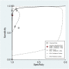
Summary ROC curve for the diagnostic performance of two-dimensional transvaginal ultrasonography to detect uterine septum, showing the sensitivity and specificity for each study and pooled estimation. The dashed line around the summary point estimate (red diamond) represents the 95% confidence region. The dotted line showing the 95% prediction contour corresponds to the predicted performance taking into account all individual studies.
Figure 5
Figure 5
Summary ROC curve for the diagnostic performance of two-dimensional sonohysterography to detect uterine septum, showing the sensitivity and specificity for each study and pooled estimation. The dashed line around the summary point estimate (red diamond) represents the 95% confidence region. The dotted line showing the 95% prediction contour corresponds to the predicted performance taking into account all individual studies.
Figure 6
Figure 6
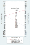
Fagan nomogram for two-dimensional transvaginal ultrasonography. It can be observed how the test changes the pre-test probability depending on a positive or negative result.
Figure 7
Figure 7
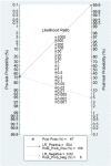
Fagan nomogram for two-dimensional transvaginal sonohysterography. It can be observed how the test changes the pre-test probability depending on a positive or negative result.
Figure 8
Figure 8
Publication bias regarding two-dimensional transvaginal ultrasonography.
Figure 9
Figure 9
Publication bias regarding two-dimensional transvaginal sonohysterography.
Figure 10
Figure 10
Forest plot for sensitivity and specificity for all studies concerning the diagnostic performance of three-dimensional transvaginal ultrasonography for uterine septum detection [13,24,25,30,34,35,38].
Figure 11
Figure 11
Summary ROC curve for the diagnostic performance of three-dimensional transvaginal ultrasonography to detect uterine septum, showing the sensitivity and specificity for each study and pooled estimation. The dashed line around the summary point estimate (red diamond) represents the 95% confidence region. The dotted line showing the 95% prediction contour corresponds to the predicted performance taking into account all individual studies.
Figure 12
Figure 12
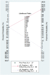
Fagan nomogram for three-dimensional transvaginal ultrasonography. It can be observed how the test changes the pre-test probability depending on a positive or negative result.
Figure 13
Figure 13
Publication bias regarding three-dimensional transvaginal ultrasonography.

References

    1. Grimbizis G.F., Campo R. Congenital Malformations of the Female Genital Tract: The Need for a New Classification System. Fertil. Steril. 2010;94:401–407. doi: 10.1016/j.fertnstert.2010.02.030. - DOI - PubMed
    1. Saravelos S.H., Cocksedge K.A., Li T.C. Prevalence and Diagnosis of Congenital Uterine Anomalies in Women with Reproductive Failure: A Critical Appraisal. Hum. Reprod. Update. 2008;14:415–429. doi: 10.1093/humupd/dmn018. - DOI - PubMed
    1. Chan Y.Y., Jayaprakasan K., Zamora J., Thornton J.G., Raine-Fenning N., Coomarasamy A. The Prevalence of Congenital Uterine Anomalies in Unselected and High-Risk Populations: A Systematic Review. Hum. Reprod. Update. 2011;17:761–771. doi: 10.1093/humupd/dmr028. - DOI - PMC - PubMed
    1. Grimbizis G.F., Camus M., Tarlatzis B.C., Bontis J.N., Devroey P. Clinical Implications of Uterine Malformations and Hysteroscopic Treatment Results. Hum. Reprod. Update. 2001;7:161–174. doi: 10.1093/humupd/7.2.161. - DOI - PubMed
    1. Tomaževič T., Ban-Frangež H., Ribič-Pucelj M., Premru-Sršen T., Verdenik I. Small Uterine Septum Is an Important Risk Variable for Preterm Birth. Eur. J. Obstet Gynecol. Reprod. Biol. 2007;135:154–157. doi: 10.1016/j.ejogrb.2006.12.001. - DOI - PubMed

LinkOut - more resources