Review
. 2023 Jan 26;10(2):218.
doi: 10.3390/children10020218.

The Use of Stress Cardiovascular Imaging in Pediatric Population

Affiliations
Review

The Use of Stress Cardiovascular Imaging in Pediatric Population

Sara Moscatelli et al. Children (Basel). .

Abstract

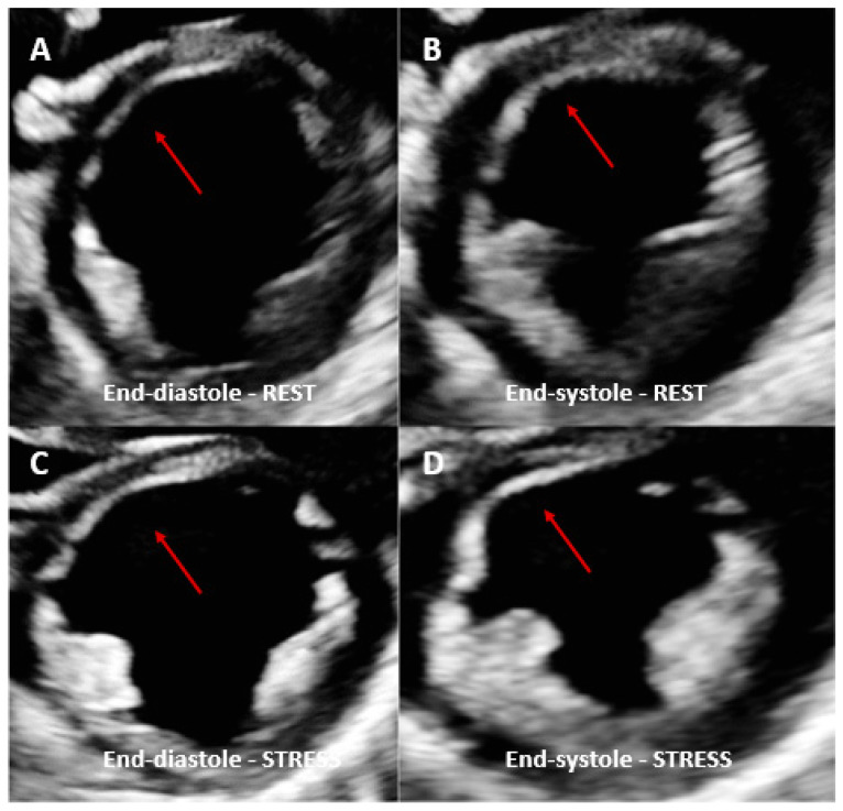
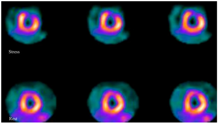
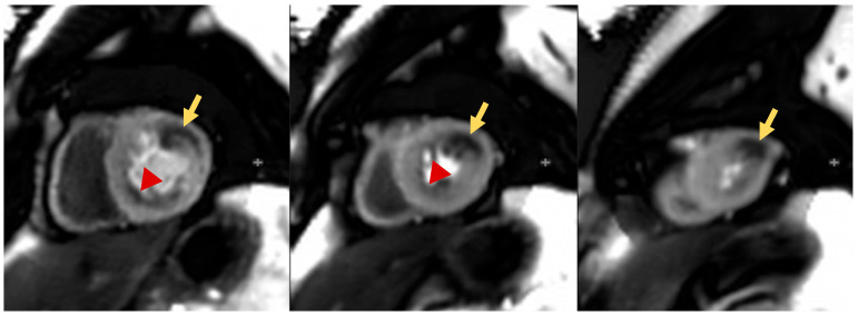
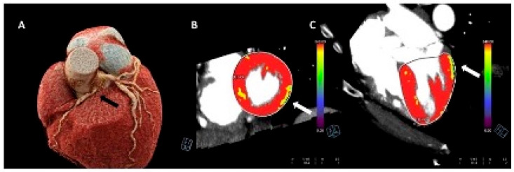
Although not frequent in the pediatric population, ischemia could occur in children due to several congenital and acquired disease. Stress imaging is key for the non-invasive evaluation of myocardial abnormalities and perfusion defect in this clinical setting. Moreover, beyond ischemia assessment, it can provide complementary diagnostic and prognostic information in valvular heart disease and cardiomyopathies. When performed using cardiovascular magnetic resonance, it could detect, in addition, myocardial fibrosis and infarction, increasing the diagnostic yield. Several imaging modalities are currently available for the evaluation of stress myocardial perfusion. Advances in technologies have also increased the feasibility, safety and availability of these modalities in the pediatric age group. However, despite the established role of stress imaging and its increasing use in daily clinical practice, there are currently no specific guidelines, and little data are available in the literature on this topic. The aim of this review is to summarize the most recent evidence on pediatric stress imaging and its clinical application with a focus on the advantages and limitations of each imaging modality currently available.

Keywords: nuclear imaging; pediatric cardiology; stress CMR; stress CT; stress cardiovascular imaging; stress echocardiography.


Conflict of interest statement

All authors declare no conflict of interest. Chiara Bucciarelli Ducci is the Chief Executive Officer (part-time) for the Society for Cardiovascular Magnetic Resonance. However, she has received speaker’s fees from Circle Cardiovascular Imaging, Bayer and Siemens Healthineers.

Figures

Figure 1
Figure 1
Dobutamine stress echo performed in a patient with coronary artery vasculopathy (CAV). End-diastolic rest images (A) demonstrated a thinned anterior septal area compared to the other myocardial segments (red arrows) and not thickening during systole (B). Same view acquired at peak dobutamine stress (C,D) demonstrated no significant changes in the same area with no other regional wall motion abnormalities identified. Angiography was therefore performed, showing severe left anterior descending coronary artery stenosis.
Figure 2
Figure 2
Axial images of a myocardial scintigraphy performed in a pediatric patient with KD: images show a severe reversible perfusion defect in the anterior wall of left ventricle, which is suggestive for myocardial ischemia.
Figure 3
Figure 3
Pediatric patients affected by KD with multiple aneurysms. The stress perfusion sequences show significant myocardial perfusion defects in the basal to mid anterolateral and apical lateral walls (likely circumflex territory, yellow arrows). Separately, there is also a mild subendocardial perfusion defect in the basal to mid inferior wall and inferoseptum (likely right coronary artery territory, red arrow heads).
Figure 4
Figure 4
Stress-computed tomography perfusion imaging of a 15-year-old boy with an anomalous origin of the right coronary artery from the left sinus of valsalva. The black arrow indicates the coronary artery abnormality (Panel (A)). The images with arrows (B,C) indicate stress-induced perfusion defects of the infero-lateral wall of the left ventricle.

References

    1. Paridon S.M., Alpert B.S., Boas S.R., Cabrera M.E., Caldarera L.L., Daniels S.R., Kimball T.R., Knilans T.K., Nixon P.A., Rhodes J., et al. Clinical Stress Testing in the Pediatric Age Group. Circulation. 2006;113:1905–1920. doi: 10.1161/CIRCULATIONAHA.106.174375. - DOI - PubMed
    1. Neumann F.J., Sousa-Uva M., Ahlsson A., Alfonso F., Banning A.P., Benedetto U., Byrne R.A., Collet J.P., Falk V., Head S.J., et al. ESC Scientific Document Group. 2018 ESC/EACTS Guidelines on myocardial revascularization. Eur. Heart J. 2019;40:87–165. doi: 10.1093/eurheartj/ehy394. - DOI - PubMed
    1. Knuuti J., Wijns W., Saraste A., Capodanno D., Barbato E., Funck-Brentano C., Prescott E., Storey R.F., Deaton C., Cuisset T., et al. 2019 ESC Guidelines for the diagnosis and management of chronic coronary syndromes. Eur. Heart J. 2020;41:407–477. doi: 10.1093/eurheartj/ehz425. - DOI - PubMed
    1. Toro-Salazar O.H., Gillan E., O’Loughlin M.T., Burke G.S., Ferranti J., Stainsby J., Liang B., Mazur W., Raman S.V., Hor K.N. Occult cardiotoxicity in childhood cancer survivors exposed to anthracycline therapy. Circ Cardiovasc. Imaging. 2013;6:873–880. doi: 10.1161/CIRCIMAGING.113.000798. - DOI - PubMed
    1. Pahl E., Duffy C.E., Chaudhry F.A. The role of stress echocardiography in children. Echocardiography. 2000;17:507–512. doi: 10.1111/j.1540-8175.2000.tb01171.x. - DOI - PubMed

LinkOut - more resources