

Muscle Activity Characteristics of the Pronator Teres during Throwing in Baseball Pitchers: A Pilot Study
- PMID: 36833152
- PMCID: PMC9957271
- DOI: 10.3390/healthcare11040618
Muscle Activity Characteristics of the Pronator Teres during Throwing in Baseball Pitchers: A Pilot Study
Abstract
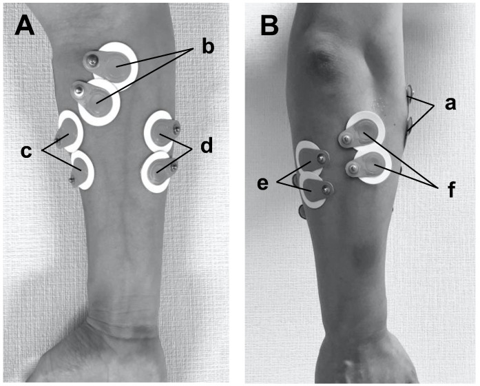
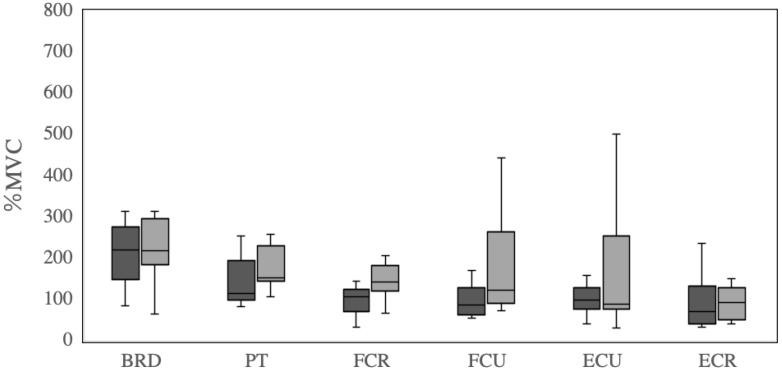
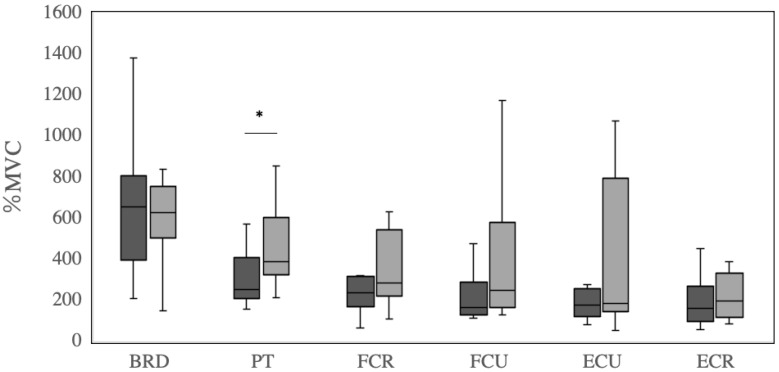
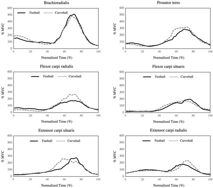
The pronator teres muscle is a major dynamic stabilizer of elbow valgus stress during throwing. This study aims to investigate pronator teres muscle activation during breaking ball pitching in baseball pitchers. Twelve male college baseball players with more than eight years of baseball experience were included in this study. A wireless surface electromyography (EMG) system was used to measure the activation of the forearm muscles and record EMG data during fastball and curveball pitching. Peak pronator teres muscle activation during curveball pitching was greater than that during fastball pitching (p = 0.03). There was no difference in the muscle activation of the other forearm muscles (p > 0.05). These results indicate that increased muscle activity in the pronator teres may contribute to stiffness and induce pronator teres syndrome or medial elbow injuries related to the overuse of the pronator teres, especially during curveball pitching. Controlling curveball throws contributes to player coaching and conditioning for the prevention of elbow joint disorders and pronator teres syndrome.
Keywords: elbow joint; electromyography; forearm; injury prevention.
Conflict of interest statement
The authors declare no conflict of interest.
Figures
Similar articles
-
A biomechanical comparison of the fastball and curveball in adolescent baseball pitchers.Am J Sports Med. 2009 Aug;37(8):1492-8. doi: 10.1177/0363546509333264. Epub 2009 May 15. Am J Sports Med. 2009. PMID: 19448049
-
Elasticity of the pronator teres muscle in youth baseball players with elbow injuries: evaluation using ultrasound strain elastography.J Shoulder Elbow Surg. 2018 Sep;27(9):1642-1649. doi: 10.1016/j.jse.2018.05.021. Epub 2018 Jun 22. J Shoulder Elbow Surg. 2018. PMID: 29941303
-
Biomechanical Comparisons Among Fastball, Slider, Curveball, and Changeup Pitch Types and Between Balls and Strikes in Professional Baseball Pitchers.Am J Sports Med. 2017 Dec;45(14):3358-3367. doi: 10.1177/0363546517730052. Epub 2017 Oct 2. Am J Sports Med. 2017. PMID: 28968139
-
The ulnar collateral ligament loading paradox between in-vitro and in-vivo studies on baseball pitching (narrative review).Int Biomech. 2021 Dec;8(1):19-29. doi: 10.1080/23335432.2021.1916405. Int Biomech. 2021. PMID: 33998377 Free PMC article. Review.
-
Baseball pitching biomechanics in relation to pain, injury, and surgery: A systematic review.J Sci Med Sport. 2021 Jan;24(1):13-20. doi: 10.1016/j.jsams.2020.06.015. Epub 2020 Jul 1. J Sci Med Sport. 2021. PMID: 32636133
Cited by
-
Diagnostic ultrasonography of upper extremity dynamic compressive neuropathies in athletes: A narrative review.Int Orthop. 2025 Apr;49(4):925-933. doi: 10.1007/s00264-025-06417-3. Epub 2025 Jan 30. Int Orthop. 2025. PMID: 39883178 Review.
References
LinkOut - more resources
Full Text Sources
