. 2023 Jan 22;14(2):291.
doi: 10.3390/genes14020291.

Stargardt-like Clinical Characteristics and Disease Course Associated with Variants in the WDR19 Gene

Affiliations

Stargardt-like Clinical Characteristics and Disease Course Associated with Variants in the WDR19 Gene

Jana Sajovic et al. Genes (Basel). .

Abstract

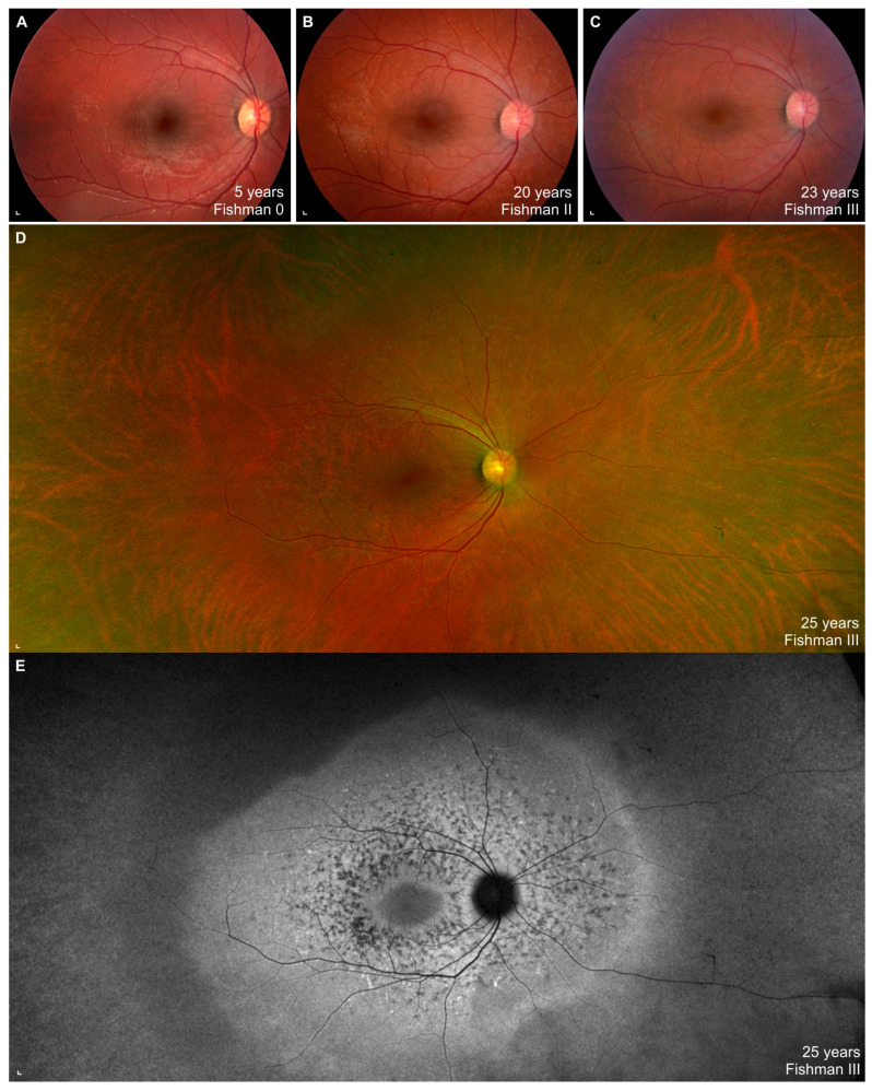
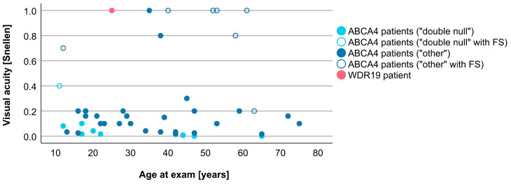
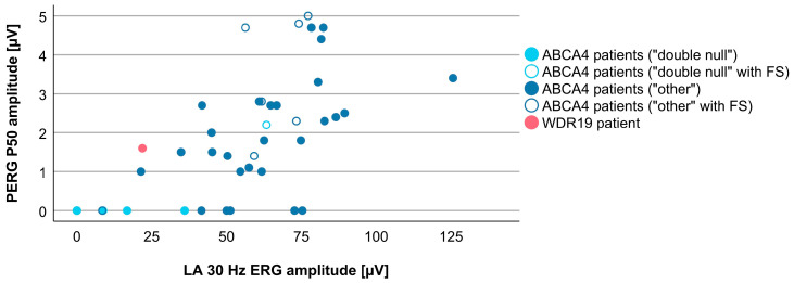
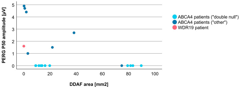
Variants in WDR19 (IFT144) have been implicated as another possible cause of Stargardt disease. The purpose of this study was to compare longitudinal multimodal imaging of a WDR19-Stargardt patient, harboring p.(Ser485Ile) and a novel c.(3183+1_3184-1)_(3261+1_3262-1)del variant, with 43 ABCA4-Stargardt patients. Age at onset, visual acuity, Ishihara color vision, color fundus, fundus autofluorescence (FAF), spectral-domain optical coherence tomography (OCT) images, microperimetry and electroretinography (ERG) were evaluated. First symptom of WDR19 patient was nyctalopia at the age of 5 years. After the age of 18 years, OCT showed hyper-reflectivity at the level of the external limiting membrane/outer nuclear layer. There was abnormal cone and rod photoreceptor function on ERG. Widespread fundus flecks appeared, followed by perifoveal photoreceptor atrophy. Fovea and peripapillary retina remained preserved until the latest exam at 25 years of age. ABCA4 patients had median age of onset at 16 (range 5-60) years and mostly displayed typical Stargardt triad. A total of 19% had foveal sparing. In comparison to ABCA4 patients, the WDR19 patient had a relatively large foveal preservation and severe rod photoreceptor impairment; however, it was still within the ABCA4 disease spectrum. Addition of WDR19 in the group of genes producing phenocopies of Stargardt disease underlines the importance of genetic testing and may help to understand its pathogenesis.

Keywords: ABCA4; IFT144; Stargardt disease; Stargardt-like disease; WDR19; fundus flavimaculaus; phenocopy.


Conflict of interest statement

The authors declare no conflict of interest.

Figures

Figure 1
Figure 1
Right eye color fundus images and fundus autofluorescence (FAF) Optos image from a WDR19 patient from the first until the last exam. Note the progression from normal fundus ((A); Fishman 0) to extensive flecks ((B); Fishman II) to resorbed flecks ((CE); Fishman III). Scale bars: 200 µm.
Figure 2
Figure 2
Right eye (left column) and left eye (right column) spectral-domain optical coherence tomography (OCT) and FAF images of WDR19 patient over the course of 7 years. Age at the exam, best-corrected visual acuity (BCVA) in Snellen, Ishihara color vision are stated on each image. Microperimetry exam is overlaid on FAF images in selected exams. Static perimetry covering central 60° degrees is shown on the bottom. Note the typical signs of Stargardt disease, i.e., peripapillary sparing and hyper-and hypoautofluorescent flecks. Macular atrophy is most notable in the perifoveal area, while fovea is preserved (i.e., foveal sparing). Inner segment ellipsoid (ISe) band is marked with arrows. Abbreviation explanation: VA—visual acuity, Ish—Ishihara plates. Scale bars: 200 µm.
Figure 3
Figure 3
Right eye (left column) and left eye (right column) electroretinography (ERG) responses of the WDR19 patient at 24 years of age (black lines) and a healthy control (grey lines). Multifocal ERG (mfERG) responses of the WDR19 patient are shown at the top with a 3D representation of the values on the right side. All ERG responses of the WDR19 patient were below the normal range. Note the relatively severe loss of ERG responses representing rod system function (dark-adapted (DA) 0.01 ERG and DA 3.0 ERG) in comparison to light-adapted (LA) 30 Hz, which represents cone system function. Preserved foveal function in the first mfERG ring is marked with green. Abbreviation explanation: PERG—pattern ERG, OP—oscillatory potentials.
Figure 4
Figure 4
Characteristics of a WDR19 patient (A) and ABCA4 patients (BF) and with foveal sparing. Note a relatively large extent of foveal preservation in the WDR19 patient. WDR19 patient is encircled with red (A), double null ABCA4 patient with light blue (B) and ABCA4 patients with other genotypes with dark blue (CF). Note the hyper-reflectivity at the level of the external limiting membrane (ELM)/outer nuclear layer (ONL) in the WDR19 patient and two ABCA4 patients (B,C). Note also the relatively dark fovea of the WDR19 patient and two of the ABCA4 patients (E,F). Scale bars: 200 µm.
Figure 5
Figure 5
Comparison of VA between WDR19 patient (red), double null ABCA4 patients (light blue) and ABCA4 patients with other genotypes (dark blue). Foveal sparing patients are marked with empty circles. Abbreviation explanation: FS—foveal sparing.
Figure 6
Figure 6
Comparison of DDAF area, representing retinal pigment epithelium (RPE) atrophy, between WDR19 patient (red), double null ABCA4 patients (light blue) and ABCA4 patients with other genotypes (dark blue). For this analysis a limited number of “other” ABCA4 patients (only those harboring ABCA4 variant c.5714+5G>A (p.[=,Glu1863Leufs*33]) was included (see Section 2).
Figure 7
Figure 7
Comparison of DA 3.0 ERG a-wave amplitudes (providing a measure of rod photoreceptor funtion) between WDR19 patient (red), double null ABCA4 patients (light blue) and ABCA4 patients with other genotypes (dark blue). Foveal sparing patients are marked with empty circles.
Figure 8
Figure 8
Comparison of DA 0.01 ERG b-wave amplitude (representing rod system function) between WDR19 patient (red), double null ABCA4 patients (light blue) and ABCA4 patients with other genotypes (dark blue). Foveal sparing patients are marked with empty circles.
Figure 9
Figure 9
Comparison of LA 30 Hz ERG amplitudes (representing cone system function) between WDR19 patient (red), double null ABCA4 patients (light blue) and ABCA4 patients with other genotypes (dark blue). Foveal sparing patients are marked with empty circles.
Figure 10
Figure 10
Comparison of PERG P50 amplitudes (representing macular function) between WDR19 patient (red), double null ABCA4 patients (light blue) and ABCA4 patients with other genotypes (dark blue). Foveal sparing patients are marked with empty circles.
Figure 11
Figure 11
Different ratios of cones and rods impairment in WDR19 patient (red), double null ABCA4 patients (light blue) and ABCA4 patients with other genotypes (dark blue). Foveal sparing patients are marked with empty circles.
Figure 12
Figure 12
Different ratios of cone system and macular photoreceptors impairment in WDR19 patient (red), double null ABCA4 patients (light blue) and ABCA4 patients with other genotypes (dark blue). Foveal sparing patients are marked with empty circles.
Figure 13
Figure 13
Different ratios of RPE and photoreceptor impairment in WDR19 patient (red), double null ABCA4 patients (light blue) and ABCA4 patients with other genotypes (dark blue). For this analysis a limited number of “other” ABCA4 patients (only those harboring ABCA4 variant c.5714+5G>A (p.[=,Glu1863Leufs * 33]) was included (see Materials and Methods).

Similar articles

Cited by

References

    1. Cremers F.P.M., Lee W., Collin R.W.J., Allikmets R. Clinical spectrum, genetic complexity and therapeutic approaches for retinal disease caused by ABCA4 mutations. Prog. Retin. Eye Res. 2020;79:100861. doi: 10.1016/j.preteyeres.2020.100861. - DOI - PMC - PubMed
    1. Fishman G.A. Fundus Flavimaculatus. A Clinical Classification. Arch. Ophthalmol. 1976;94:2061–2067. doi: 10.1001/archopht.1976.03910040721003. - DOI - PubMed
    1. Tanna P., Strauss R.W., Fujinami K., Michaelides M. Stargardt disease: Clinical features, molecular genetics, animal models and therapeutic options. Br. J. Ophthalmol. 2017;101:25–30. doi: 10.1136/bjophthalmol-2016-308823. - DOI - PMC - PubMed
    1. Molday L.L., Rabin A.R., Molday R.S. ABCR expression in foveal cone photoreceptors and its role in Stargardt macular dystrophy. Nat. Genet. 2000;25:257–258. doi: 10.1038/77004. - DOI - PubMed
    1. Illing M., Molday L.L., Molday R.S. The 220-kDa Rim Protein of Retinal Rod Outer Segments Is a Member of the ABC Transporter Superfamily. J. Biol. Chem. 1997;272:10303–10310. doi: 10.1074/jbc.272.15.10303. - DOI - PubMed

Publication types

Substances

LinkOut - more resources