. 2023 Jan 25;14(2):309.
doi: 10.3390/genes14020309.

Characterization and Expression of Holothurian Wnt Signaling Genes during Adult Intestinal Organogenesis

Affiliations

Characterization and Expression of Holothurian Wnt Signaling Genes during Adult Intestinal Organogenesis

Noah A Auger et al. Genes (Basel). .

Abstract

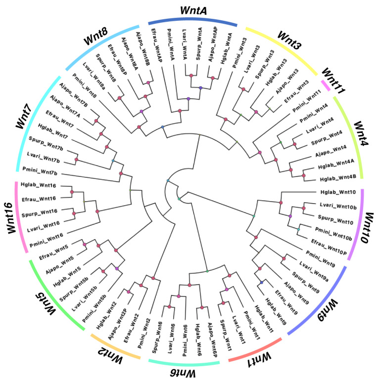
Wnt signaling has been shown to play multiple roles in regenerative processes, one of the most widely studied of which is the regeneration of the intestinal luminal epithelia. Most studies in this area have focused on self-renewal of the luminal stem cells; however, Wnt signaling may also have more dynamic functions, such as facilitating intestinal organogenesis. To explore this possibility, we employed the sea cucumber Holothuria glaberrima that can regenerate a full intestine over the course of 21 days after evisceration. We collected RNA-seq data from various intestinal tissues and regeneration stages and used these data to define the Wnt genes present in H. glaberrima and the differential gene expression (DGE) patterns during the regenerative process. Twelve Wnt genes were found, and their presence was confirmed in the draft genome of H. glaberrima. The expressions of additional Wnt-associated genes, such as Frizzled and Disheveled, as well as genes from the Wnt/β-catenin and Wnt/Planar Cell Polarity (PCP) pathways, were also analyzed. DGE showed unique distributions of Wnt in early- and late-stage intestinal regenerates, consistent with the Wnt/β-catenin pathway being upregulated during early-stages and the Wnt/PCP pathway being upregulated during late-stages. Our results demonstrate the diversity of Wnt signaling during intestinal regeneration, highlighting possible roles in adult organogenesis.

Keywords: Wnt genes; echinoderm; organogenesis; regeneration; sea cucumber.


Conflict of interest statement

The authors declare no conflict of interest.

Figures

Figure 1
Figure 1
The process of intestinal organogenesis after evisceration. Beneath each timepoint is a color-coded cellular process unique to either the mesothelium (light blue) or luminal epithelium (dark pink). Normal Intestine: a normal intestine connected to the body wall with the mesentery and intestine distinguished, which are the controls for the rudiment and lumen, respectively. 0-hpe: the eviscerated intestine leaves a torn edge at the mesenterial tip, which is the site of injury response and initial wound healing, thus beginning the process of regeneration. 12-hpe: dedifferentiation occurs in the mesothelium, which is composed of peritoneocytes and myoepithelium; this process is characterized by the presence of spindle like structures that are discarded actin filaments from myoepithelial cells. 1-dpe: dedifferentiation of the mesothelium continues in a gradient along the mesentery, moving proximally toward the body wall. 3-dpe: some dedifferentiated cells ingress into the connective tissue (CT) of the rudiment, undergoing an epithelial-to-mesenchymal transition (EMT). 7-dpe: proliferation in the regenerating rudiment picks up pace; simultaneously, a few cells begin redifferentiating into mature mesothelium. 14-dpe: the rudiment is invaded by luminal epithelium derived from the esophagus (anterior) and the cloaca (posterior) that extend toward one another. The dashed line shows the anterior and posterior intestine with luminal epithelium and a surrounding mesenchyme (above dash), while in between is the rudiment without a lumen (below dash). 21-dpe: the anterior and posterior luminal epithelium connect to form a continuous lumen.
Figure 2
Figure 2
Structure of annotated Wnt family genes in H. glaberrima. The structure for all Wnt genes was characterized utilizing Wnt transcripts identified in the sea cucumber transcriptome data. All the scaffolds that contain each Wnt exon are identified by labels on top of each bracket (e.g., Hglab.02944). The start and end coordinates of the Wnts span across each scaffold are shown at the bottom of each gene structure based on the genome nucleotide sequences (NCBI ID: GCA_009936505.2). Broken lines between exons represent a gene structure break due to genome fragmentation. Exon and intron sizes are based on base pair lengths, in which each base pair is equal to 0.2 for exons and 0.05 for introns.
Figure 3
Figure 3
Phylogenetic analysis of Wnt from distinct Echinoderm species. Gene tree of all Wnt genes identified in H. glaberrima (Hglab), along with those deposited in NCBI or Echinobase for S. purpuratus (Spur), L. variegatus (Lvari), P. miniata (Pmini), A. japonicus (Ajapo), and E. fraudatrix (Efrau). For all partial sequences, a letter P was added to its label; all other letters are based on the gene name shown in NCBI. The size and color of circles in nodes are dependent on their bootstrap value. Nodes with bootstrap values over 95 are shown in pink.
Figure 4
Figure 4
Wnt cluster conservation. Schematic illustrating the conservation of the Wnt1-Wnt6-Wnt10 cluster in H. glaberrima compared to L. variegatus and Drosophila melanogaster (adapted from [47]). For H. glaberrima, we show the distribution of these genes across the draft assembly by complete squares with arrow heads pointing in the direction of the gene in the scaffold. The coordinates on top of the gene structures show scaffold size. The IDs on top of each gene structure represent the scaffold ID (NCBI ID: GCA_009936505.2). Separations of each Wnt due to fragmentation are shown as black line breaks. Start and stop codons are represented by green circles and red stars, respectively. The cluster of L. variegatus was characterized using its latest genome (Lvar_3.0; NCBI ID: 3495).
Figure 5
Figure 5
Heatmap of Wnt expression during early- and late-stage regeneration. This heatmap contains all the RNA-seq timepoints collected from our transcriptomic databases. The timepoints 12-hpe through 14-dpe should be referenced to Normal Mesentery, as this was the control. Similarly, the timepoints 14-dpeA, 14-dpeP, and 21-dpe should be referenced to Normal Intestine. On either side of the heatmap are the Log2foldchange (L2FC) values of a Wnt gene at a given timepoint. A significance threshold was set at L2FC <−2 or >2 with a pADJ value of 0.001. Above the columns are color coded labels that represent the tissue composition of the samples at a given timepoint. Abbreviations—CT: connective tissue, ME: mesothelium, and LE: luminal epithelium.
Figure 6
Figure 6
Heatmap of Fzd and Dvl expression during early- and late-stage regeneration. This figure can be read the same as the heatmap from Figure 5. The significance threshold is the same.
Figure 7
Figure 7
Heatmap of the signaling genes in the Wnt/β-catenin pathway. This figure is read the same as in Figure 5. The significance threshold remains the same. The only difference is that the rows are color coded to indicate the role of the gene in the Wnt/B-catenin pathway. Genes in the Off-state inhibit β-catenin signaling, while genes in the On-state facilitate Wnt/β-catenin signaling. The Target Genes are the downstream targets of the TCF/LEF transcription factor family that is turned on by β-catenin.
Figure 8
Figure 8
Heatmap of the signaling genes in the Wnt/PCP pathway. This figure is read the same as in Figure 5. The significance threshold remains the same. The only difference is that the rows are color coded to indicate the role of the gene in the Wnt/PCP pathway. Genes labeled as Core Proteins are unique and essential to the Wnt/PCP pathway and, therefore, are strong indicators of its presence. The effector proteins are genes involved in carrying out the signaling cascade, while target genes are activated by the transcriptions factor c-Jun.
Figure 9
Figure 9
Summary of Wnt gene compositions of the echinoderm species assessed in this study. This schematic is based on a phylogenetic analysis of Figure 2. Asterisks depict manually characterized genes. White X depicts genes that could not be found in the NCBI database.

Similar articles

Cited by

References

    1. Yaglova N.V., Tsomartova D.A., Obernikhin S.S., Nazimova S.V. The Role of the Canonical Wnt-Signaling Pathway in Morphogenesis and Regeneration of the Adrenal Cortex in Rats Exposed to the Endocrine Disruptor Dichlorodiphenyltrichloroethane during Prenatal and Postnatal Development. Biol. Bull. Russ. Acad. Sci. 2019;46:74–81. doi: 10.1134/S1062359018060122. - DOI
    1. Komiya Y., Habas R. Wnt Signal Transduction Pathways. Organogenesis. 2008;4:68–75. doi: 10.4161/org.4.2.5851. - DOI - PMC - PubMed
    1. Otto A., Schmidt C., Luke G., Allen S., Valasek P., Muntoni F., Lawrence-Watt D., Patel K. Canonical Wnt Signalling Induces Satellite-Cell Proliferation during Adult Skeletal Muscle Regeneration. J. Cell Sci. 2008;121:2939–2950. doi: 10.1242/jcs.026534. - DOI - PubMed
    1. He T.-C., Sparks A.B., Rago C., Hermeking H., Zawel L., da Costa L.T., Morin P.J., Vogelstein B., Kinzler K.W. Identification of C-MYC as a Target of the APC Pathway. Science. 1998;281:1509–1512. doi: 10.1126/science.281.5382.1509. - DOI - PubMed
    1. Tetsu O., McCormick F. B-Catenin Regulates Expression of Cyclin D1 in Colon Carcinoma Cells. Nature. 1999;398:422–426. doi: 10.1038/18884. - DOI - PubMed

Publication types

LinkOut - more resources