Case Reports
. 2023 Feb 6;20(4):2843.
doi: 10.3390/ijerph20042843.

Vertical Transmission of Coxsackievirus A6 with Severe Congenital Pneumonia/Sepsis

Affiliations
Case Reports

Vertical Transmission of Coxsackievirus A6 with Severe Congenital Pneumonia/Sepsis

Ruka Nakasone et al. Int J Environ Res Public Health. .

Abstract

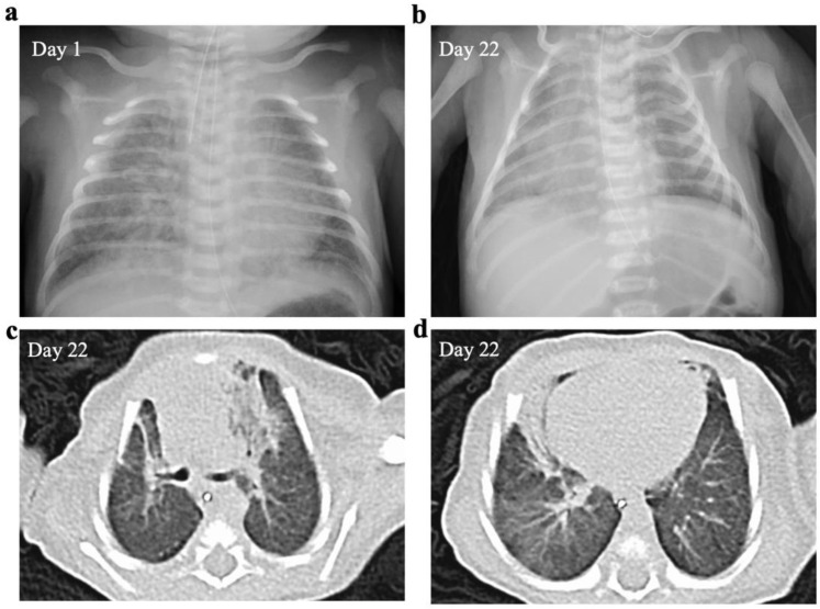
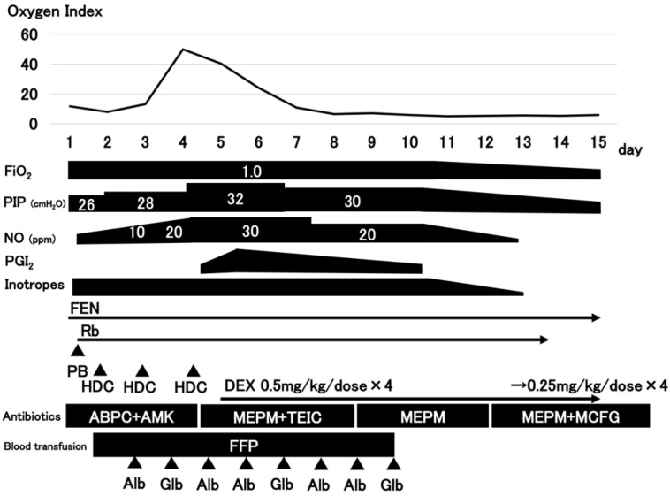
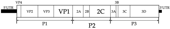
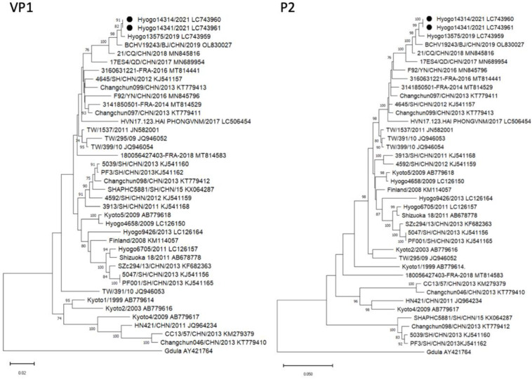
We report a case of vertical transmission of Coxsackievirus (CV)-A6 with severe congenital pneumonia/sepsis. A male infant presented with severe respiratory symptoms at birth and was treated with full cardiopulmonary support, including inhaled nitric oxide. Three days before delivery, his older brother was diagnosed with hand, foot, and mouth disease (HFMD). His mother developed transient fever 1 day before delivery and presented a blister on her thumb 2 days after delivery. A multiplex polymerase chain reaction test on day 2 was positive for human rhinovirus/enterovirus. CV-A6 was later detected in the serum, tracheal aspirate, and stool of the patient sampled on day 6, and in the maternal serum sampled on the day of delivery. He was diagnosed with congenital CV-A6 pneumonia/sepsis caused by vertical transmission, based on VP1 consensus sequences used for typing of the virus that demonstrated a 100% match between the mother and infant. Further, the strain was closely related to the lethal CV-A6-Changchun strains in the phylogenetic analysis of the P2 region, which contributes to the pathogenicity. In conclusion, congenital CV-A6 infection should be considered if a woman exhibits HFMD symptoms during the perinatal period. Detailed virologic examination is useful for understanding its pathogenesis.

Keywords: Coxsackievirus A6; HFMD; congenital pneumonia; multiplex PCR; vertical transmission.


Conflict of interest statement

The authors declare no conflict of interest.

Figures

Figure 1
Figure 1
Chest radiograph and computed tomography (CT) scan. The chest radiograph displays bilateral hazy opacities (a). The chest radiograph (b) and CT examination (c,d) performed at 22 days of age reveal infiltrative shadows in both lung fields and decreased lung capacity.
Figure 2
Figure 2
The clinical course of the patient. FiO2, fraction of inspired oxygen; PIP, peak inspiratory pressure; NO, nitric oxide; PGI2, prostaglandin I2 (epoprostenol); Inotropes (DOA, dopamine; DOB, dobutamine); FEN, fentanyl; Rb, rocuronium bromide; PB, phenobarbital; HDC, hydrocortisone; DEX, dexamethasone; ABPC, ampicillin; AMK, amikacin; MEPM, meropenem; TEIC, teicoplanin; MCFG, micafungin; FFP, fresh frozen plasma; Alb, albumin; Glb, globulin.
Figure 3
Figure 3
Structure of the enterovirus genome. The genome contains a single open reading frame that encodes a polyprotein and is flanked by 5′ and 3′ untranslated regions (UTRs). The encoded polyprotein contains three major regions (P1, P2, and P3). These regions can self-digest into four separate structural proteins (VP1, VP2, VP3, and VP4 from P1) and seven nonstructural proteins (2A, 2B, and 2C from P2; 3A, 3B, 3C, and 3D from P3) [5].
Figure 4
Figure 4
Phylogenetic analysis of the VP1 and P2 regions of the CV-A6 strains. (Left) Neighbor-joining tree based on the VP1 sequences (positions 2441 to 3355 correspond to the Gdula genome). (Right) Neighbor-joining tree based on the P2 sequences (positions 3356 to 5089 correspond to the Gdula genome). Both phylogenetic trees were tested using the bootstrap method for 1000 replicates, and values of >70 are shown at the nodes. ●, CV-A6 Hyogo strains of this case.

References

    1. Sandoni M., Ciardo L., Tamburini C., Boncompagni A., Rossi C., Guidotti I., Garetti E., Lugli L., Iughetti L., Berardi A. Enteroviral Infections in the First Three Months of Life. Pathogens. 2022;11:60. doi: 10.3390/pathogens11010060. - DOI - PMC - PubMed
    1. Yoshitomi H., Ashizuka Y., Ichihara S., Nakamura T., Nakamura A., Kobayashi T., Kajiwara J. Molecular epidemiology of coxsackievirus A6 derived from hand, foot, and mouth disease in Fukuoka between 2013 and 2017. J. Med. Virol. 2018;90:1712–1719. doi: 10.1002/jmv.25250. - DOI - PubMed
    1. Bruning A.H., van der Sanden S.M., ten Hoedt A.E., Wolthers K.C., van Kaam A.H., Pajkrt D. An atypical course of coxsackievirus A6 associated hand, foot and mouth disease in extremely low birth weight preterm twins. J. Clin. Virol. 2015;65:20–22. doi: 10.1016/j.jcv.2015.01.020. - DOI - PubMed
    1. Xu S., Li H., Qiao P., Xu G., Zhao D., Lin X., Qin Y., Yu H., Zhang X., Zhang W., et al. Neonatal hand, foot, and mouth disease due to coxsackievirus A6 in Shanghai. BMC Pediatr. 2020;20:364. doi: 10.1186/s12887-020-02262-y. - DOI - PMC - PubMed
    1. Wang S.H., Wang A., Liu P.P., Zhang W.Y., Du J., Xu S., Liu G.C., Zheng B.S., Huan C., Zhao K., et al. Divergent Pathogenic Properties of Circulating Coxsackievirus A6 Associated with Emerging Hand, Foot, and Mouth Disease. J. Virol. 2018;92:e00303–e00318. doi: 10.1128/JVI.00303-18. - DOI - PMC - PubMed

Publication types

Substances