Review
. 2023 Feb 4;24(4):3086.
doi: 10.3390/ijms24043086.

Multiplex Immunofluorescence: A Powerful Tool in Cancer Immunotherapy

Affiliations
Review

Multiplex Immunofluorescence: A Powerful Tool in Cancer Immunotherapy

Wenjie Sheng et al. Int J Mol Sci. .

Abstract

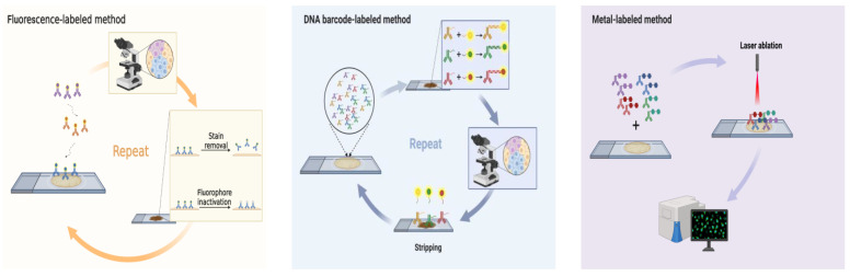
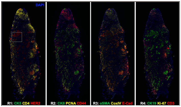
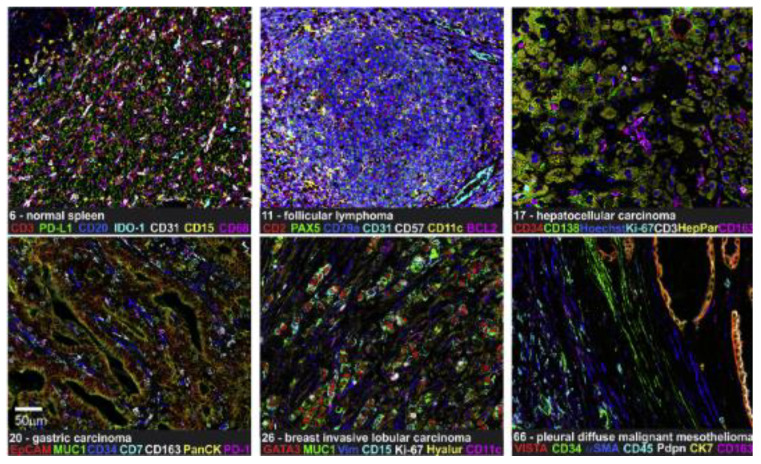
Traditional immunohistochemistry (IHC) has already become an essential method of diagnosis and therapy in cancer management. However, this antibody-based technique is limited to detecting a single marker per tissue section. Since immunotherapy has revolutionized the antineoplastic therapy, developing new immunohistochemistry strategies to detect multiple markers simultaneously to better understand tumor environment and predict or assess response to immunotherapy is necessary and urgent. Multiplex immunohistochemistry (mIHC)/multiplex immunofluorescence (mIF), such as multiplex chromogenic IHC and multiplex fluorescent immunohistochemistry (mfIHC), is a new and emerging technology to label multiple biomarkers in a single pathological section. The mfIHC shows a higher performance in cancer immunotherapy. This review summarizes the technologies, which are applied for mfIHC, and discusses how they are employed for immunotherapy research.

Keywords: antibody; cancer; fluorescence; immunohistochemistry; immunotherapy; multiplex.


Conflict of interest statement

The authors declare no conflict of interest.

Figures

Figure 4
Figure 4
Opal™ shows multiplex staining in human pancreatic ductal adenocarcinoma (PDAC) tissue sections. CK8 (green), Collagen-I (extracellular teal blue), aSMA (cytoplasmic, red), CD31 (membrane, cyan), CD4 (membrane, pseudocolored magenta), CD8 (membrane, Cy5, orange), Foxp3 (nuclear, white), autofluorescence (black), and the DAPI nuclear marker (blue). Scale bar 100 µM. (Reprinted with permission from [46], Copyright 2017 Springer Nature under a Creative Commons Attribution 4.0 International License).
Figure 6
Figure 6
IMC shows the structural and cellular iTME constitutions in the cutaneous squamous cell carcinomas (cSCC), representing lymphatic vessels (podoplanin), blood vessels and cancer-associated fibroblasts (αSMA), nerve fibers (pan-neurofilament), tumor cells (pan-cytokeratin), extracellular matrix (fibronectin) and immune cells (CD45), nuclei, and hematoxylin-eosin-saffron (HES) also were done in the same region. Scale bar 100 µm. (Reprinted with permission from [75]. Copyright 2021 Frontiers Media SA).
Figure 1
Figure 1
Schematic diagram showing the mechanism of each multiplex fluorescent immunohistochemistry method and was created with BioRender.com. Fluorescence-labeled method: multiepitope-ligand cartography (MELC), sequential immuno-peroxidase labelling and erasing (SIMPLE), iterative bleaching extends multiplexity (IBEX), multiplexed fluorescence microscopy (MxIF), cyclic immunofluorescence (CycIF), ChipCytometry, UltraPlex™, Opal™, quantum dots (QDs). DNA barcode-labeled method: DNA Exchange Imaging (DEI), codetection by indexing (CODEX), Immuno-SABER, Digital Spatial Profiling (DSP), InSituPlex®. Metal-labeled method: imaging Mass Cytometry (IMC), multiplexed ion beam imaging (MIBI). Created with BioRender.com.
Figure 2
Figure 2
HNSCC FFPE sections stained with lymphoid biomarkers. Biomarkers and colors are shown on the right. Scale bar, 500 µm. (Reprinted with permission from [24]. Copyright 2017 Elsevier).
Figure 3
Figure 3
CycIF shows whole HER2 positive breast cancer tissue images. CK5, cytokeratin 5; CK8, cytokeratin 8; CK19, cytokeratin 19; PCNA, proliferating cell nuclear antigen; E-Cad, E-cadherin; α-SMA, α-smooth muscle antigen; CoxIV, cytochrome c oxidase. (Reprinted with permission from [36]. Copyright 2020 International Society for Optical Engineering).
Figure 5
Figure 5
CODEX shows 7-plex imaging in different FFPE tissues. IDO-1, indoleamine 2,3-dioxygenase 1; Vim, vimentin; VISTA, V domain immunoglobulin suppressor of T-cell activation. Scale bar 50 µm. (Reprinted with permission from [55]. Copyright © 2020, Elsevier under Creative Commons CC-BY license).
Figure 7
Figure 7
Strengths, Weaknesses, Opportunities, and Threats (SWOT) analysis regarding mfIHC applications in clinical cancer immunotherapy.

References

    1. Coons A.H., Creech H.J., Jones R.N. Immunological properties of an antibody containing a fluorescent group. Proc. Soc. Exp. Biol. Med. 1941;47:200–202. doi: 10.3181/00379727-47-13084P. - DOI
    1. Roach C., Zhang N., Corigliano E., Jansson M., Toland G., Ponto G., Dolled-Filhart M., Emancipator K., Stanforth D., Kulangara K. Development of a Companion Diagnostic PD-L1 Immunohistochemistry Assay for Pembrolizumab Therapy in Non-Small-cell Lung Cancer. Appl. Immunohistochem. Mol. Morphol. 2016;24:392–397. doi: 10.1097/PAI.0000000000000408. - DOI - PMC - PubMed
    1. Lechner M.G., Karimi S.S., Barry-Holson K., Angell T.E., Murphy K.A., Church C.H., Ohlfest J.R., Hu P., Epstein A.L. Immunogenicity of murine solid tumor models as a defining feature of in vivo behavior and response to immunotherapy. J. Immunother. 2013;36:477–489. doi: 10.1097/01.cji.0000436722.46675.4a. - DOI - PMC - PubMed
    1. Paret C., Simon P., Vormbrock K., Bender C., Kolsch A., Breitkreuz A., Yildiz O., Omokoko T., Hubich-Rau S., Hartmann C., et al. CXorf61 is a target for T cell based immunotherapy of triple-negative breast cancer. Oncotarget. 2015;6:25356–25367. doi: 10.18632/oncotarget.4516. - DOI - PMC - PubMed
    1. Harel M., Ortenberg R., Varanasi S.K., Mangalhara K.C., Mardamshina M., Markovits E., Baruch E.N., Tripple V., Arama-Chayoth M., Greenberg E., et al. Proteomics of Melanoma Response to Immunotherapy Reveals Mitochondrial Dependence. Cell. 2019;179:236–250.e218. doi: 10.1016/j.cell.2019.08.012. - DOI - PMC - PubMed