. 2023 Feb 4;24(4):3092.
doi: 10.3390/ijms24043092.

Myxinidin-Derived Peptide against Biofilms Caused by Cystic Fibrosis Emerging Pathogens

Affiliations

Myxinidin-Derived Peptide against Biofilms Caused by Cystic Fibrosis Emerging Pathogens

Rosa Bellavita et al. Int J Mol Sci. .

Abstract

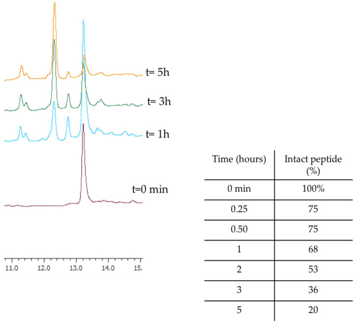
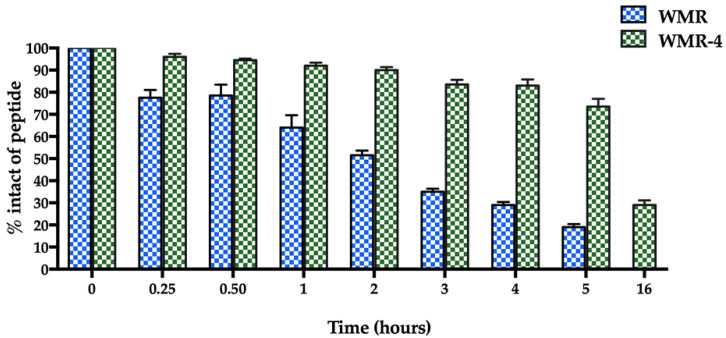
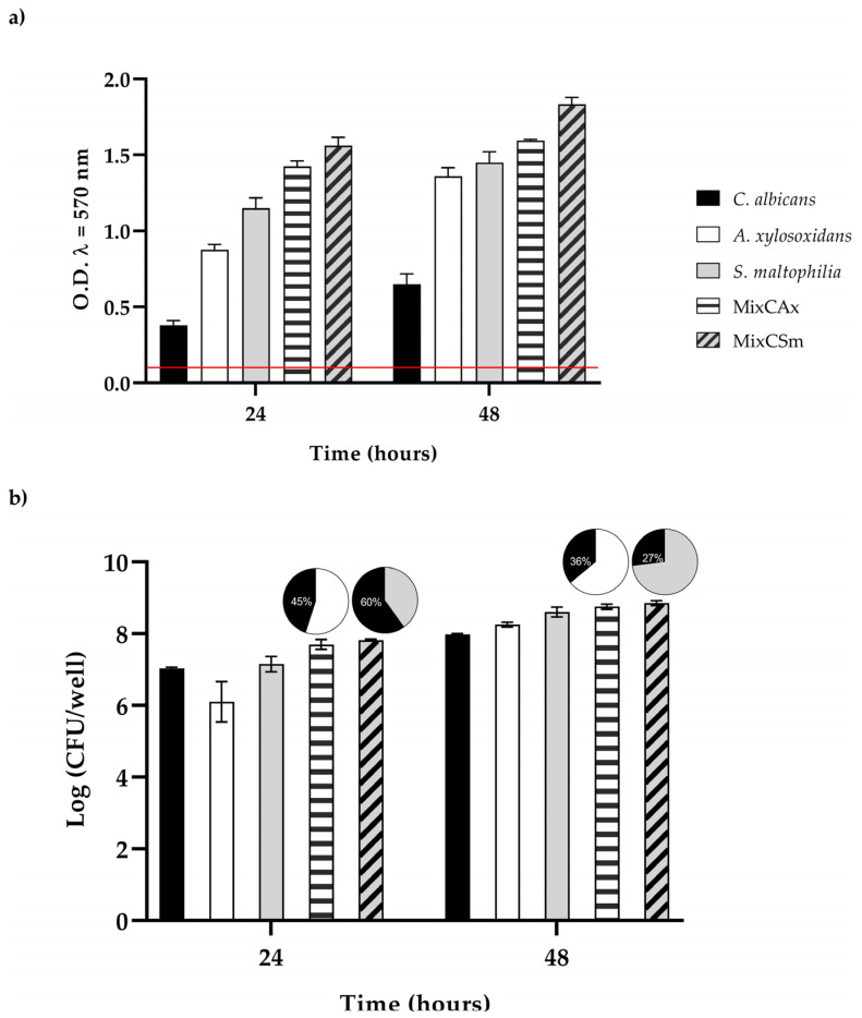
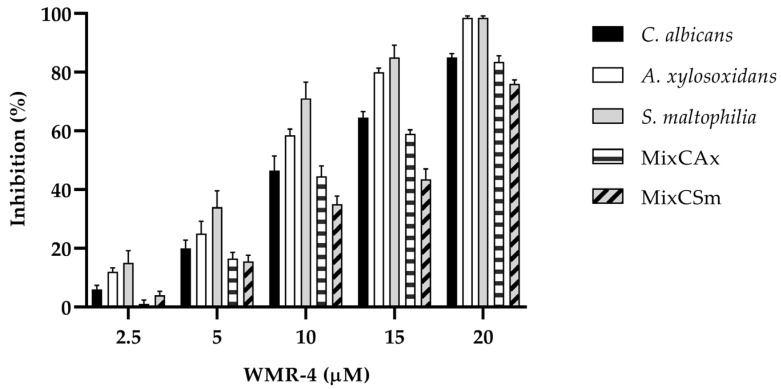
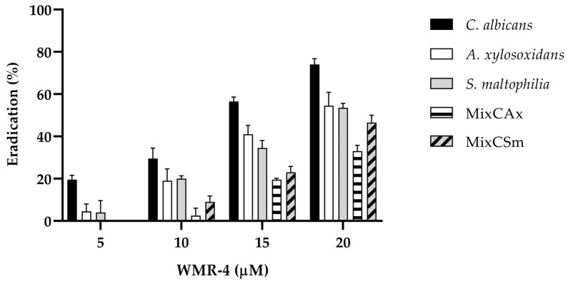
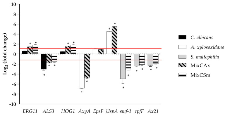
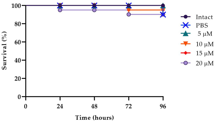
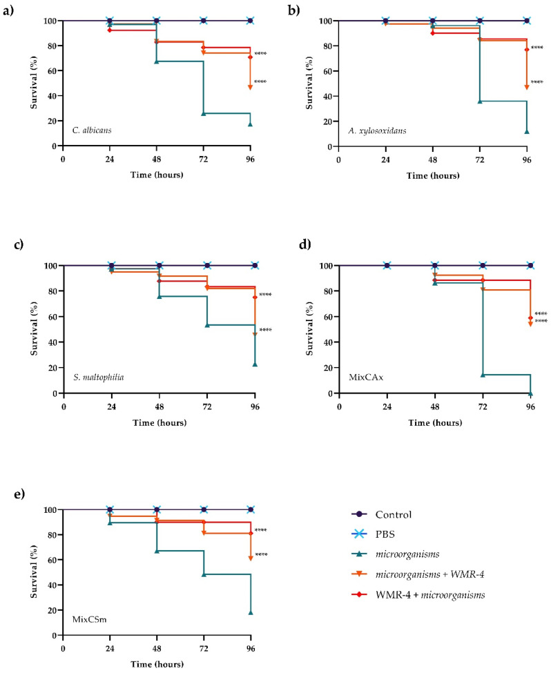
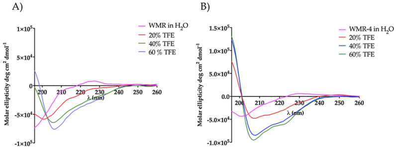
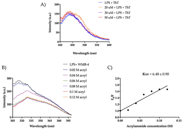
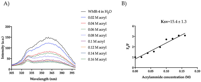
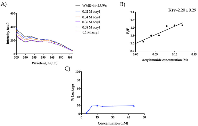
Chronic lung infections in cystic fibrosis (CF) patients are triggered by multidrug-resistant bacteria such as Pseudomonas aeruginosa, Achromobacter xylosoxidans, and Stenotrophomonas maltophilia. The CF airways are considered ideal sites for the colonization and growth of bacteria and fungi that favor the formation of mixed biofilms that are difficult to treat. The inefficacy of traditional antibiotics reinforces the need to find novel molecules able to fight these chronic infections. Antimicrobial peptides (AMPs) represent a promising alternative for their antimicrobial, anti-inflammatory, and immunomodulatory activities. We developed a more serum-stable version of the peptide WMR (WMR-4) and investigated its ability to inhibit and eradicate C. albicans, S. maltophilia, and A. xylosoxidans biofilms in both in vitro and in vivo studies. Our results suggest that the peptide is able better to inhibit than to eradicate both mono and dual-species biofilms, which is further confirmed by the downregulation of some genes involved in biofilm formation or in quorum-sensing signaling. Biophysical data help to elucidate its mode of action, showing a strong interaction of WMR-4 with lipopolysaccharide (LPS) and its insertion in liposomes mimicking Gram-negative and Candida membranes. Our results support the promising therapeutic application of AMPs in the treatment of mono- and dual-species biofilms during chronic infections in CF patients.

Keywords: Achromobacter xylosoxidans; Candida albicans; Stenotrophomonas maltophilia; antibiofilm activity; antimicrobial peptides; cystic fibrosis; membrane interaction; polymicrobial infections.


Conflict of interest statement

The authors declare no conflict of interest.

Figures

Figure 1
Figure 1
The representative RP-HPLC chromatograms of WMR at different times (0, 1, 3, and 5 h), and the percentage of intact peptide after incubation with 50% bovine serum at 37 °C.
Figure 2
Figure 2
The serum stability of peptides WMR and WMR-4 in bovine serum determined by RP-HPLC. The percentage of intact peptide was obtained by calculating the relative peak area monitored by RP-HPLC and subtracting background peak areas in blank matrix.
Figure 3
Figure 3
(a) Biofilm formation capacity of C. albicans, A. xylosoxidans, S. maltophilia, and the two mixed biofilms (CAx, CSm) using the crystal violet staining method. Red line represents ODcut. ODcut = mean of negative control with addition of 3 times the SD. (b) Enumeration of colony forming units per well for single- and dual-species biofilms.
Figure 4
Figure 4
Antibiofilm activity of WMR-4 on C. albicans, S. maltophilia, A. xylosoxidans, and two mixed biofilms (CAx, CSm) quantified with crystal violet after 24 h.
Figure 5
Figure 5
Disruption of established biofilms of C. albicans, S. maltophilia, and A. xylosoxidans and two mixed biofilms (CAx, CSm) after the treatment with the peptide WMR-4.
Figure 6
Figure 6
Real-time qPCR during inhibition of single and mixed biofilm using WMR-4 at concentration of 10 µM. Histograms represent the fold differences in the expression levels of the genes selected during inhibition of single and mixed biofilm with WMR-4 at concentration of 10 µM. Red lines show fold change thresholds of −1 and +1, respectively. * = p < 0.05.
Figure 7
Figure 7
Peptide toxicity on G. mellonella larvae treated with WMR-4 at the concentrations of 5 μM, 10 μM, 15 μM, and 20 μM.
Figure 8
Figure 8
Kaplan–Meier plots of survival curves of G. mellonella larvae infected with C. albicans (a), A. xylosoxidans (b), S. maltophilia (c), CAx (d), CSm (e). The concentration of microorganisms was 1 × 10⁶ CFU/larva. All groups were treated with 10 μM WMR-4 before or after infection/co-infection. All groups were compared with control (infected or co-infected larvae). In all panels, survival curves of intact larvae and larvae treated with PBS are reported. **** Represents p-value < 0.001 (Tukey’s).
Figure 9
Figure 9
CD spectra of peptides WMR (A) and WMR-4 (B) in water and in presence of 20%, 40%, and 60% of TFE.
Figure 10
Figure 10
(A) ThT emission spectra after the incubation of WMR-4 (20, 30, and 50 μM) with LPS. (B) Tryptophan fluorescence spectra for the peptide WMR-4 in LPS during the quenching with acrylamide at different concentrations. (C) Stern–Volmer (Ksv) quenching constant of WMR-4 in the presence of LPS.
Figure 11
Figure 11
Tryptophan fluorescence spectra for the peptide WMR-4 in water during the quenching with acrylamide at different concentrations (A) and Stern–Volmer (Ksv) quenching constant (B).
Figure 12
Figure 12
Tryptophan fluorescence spectra for the peptide WMR-4 in LUVs mimicking Gram-negative membranes during the quenching with acrylamide at different concentrations (A) and Stern–Volmer (Ksv) quenching constant (B). The leakage percentage of LUVs (DOPE/DOPG/CL) induced by WMR-4 (C).
Figure 13
Figure 13
Trp fluorescence spectra for the peptide WMR-4 in LUVs of PE/PC/PI/Erg (A) and Ksv quenching constant (B).

References

    1. Allan K.M., Farrow N., Donnelley M., Jaffe A., Waters S.A. Treatment of cystic fibrosis: From gene- to cell-based therapies. Front. Pharmacol. 2021;12:639475. doi: 10.3389/fphar.2021.639475. - DOI - PMC - PubMed
    1. Boucher R.C. New concepts of the pathogenesis of cystic fibrosis lung disease. Eur. Respir. J. 2004;23:146–158. doi: 10.1183/09031936.03.00057003. - DOI - PubMed
    1. Cutting G.R. Cystic fibrosis genetics: From molecular understanding to clinical application. Nat. Rev. Genet. 2015;16:45–56. doi: 10.1038/nrg3849. - DOI - PMC - PubMed
    1. Valenza G., Tappe D., Turnwald D., Frosch M., König C., Hebestreit H., Abele-Horn M. Prevalence and antimicrobial susceptibility of microorganisms isolated from sputa of patients with cystic fibrosis. J. Cyst. Fibros. 2008;7:123–127. doi: 10.1016/j.jcf.2007.06.006. - DOI - PubMed
    1. Kreda S.M., Davis C.W., Rose M.C. CFTR, mucins, and mucus obstruction in cystic fibrosis. Cold Spring Harb. Perspect. Med. 2012;2:a009589. doi: 10.1101/cshperspect.a009589. - DOI - PMC - PubMed

LinkOut - more resources