

Protective Barriers Provided by the Epidermis
- PMID: 36834554
- PMCID: PMC9961209
- DOI: 10.3390/ijms24043145
Protective Barriers Provided by the Epidermis
Abstract
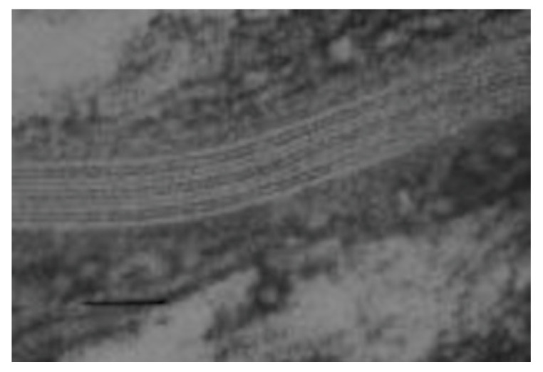
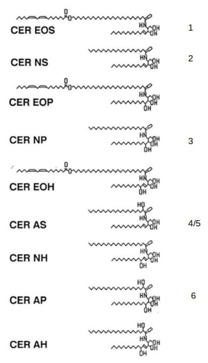
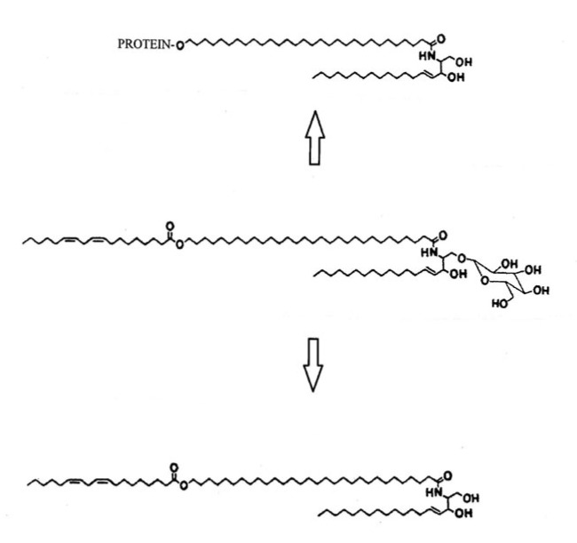
The skin is the largest organ of the body and consists of an epidermis, dermis and subcutaneous adipose tissue. The skin surface area is often stated to be about 1.8 to 2 m2 and represents our interface with the environment; however, when one considers that microorganisms live in the hair follicles and can enter sweat ducts, the area that interacts with this aspect of the environment becomes about 25-30 m2. Although all layers of the skin, including the adipose tissue, participate in antimicrobial defense, this review will focus mainly on the role of the antimicrobial factors in the epidermis and at the skin surface. The outermost layer of the epidermis, the stratum corneum, is physically tough and chemically inert which protects against numerous environmental stresses. It provides a permeability barrier which is attributable to lipids in the intercellular spaces between the corneocytes. In addition to the permeability barrier, there is an innate antimicrobial barrier at the skin surface which involves antimicrobial lipids, peptides and proteins. The skin surface has a low surface pH and is poor in certain nutrients, which limits the range of microorganisms that can survive there. Melanin and trans-urocanic acid provide protection from UV radiation, and Langerhans cells in the epidermis are poised to monitor the local environment and to trigger an immune response as needed. Each of these protective barriers will be discussed.
Keywords: Langerhans cells; antimicrobial lipids; antimicrobial peptides; barrier function; epidermis; melanocytes.
Conflict of interest statement
The authors have no conflict of interest.
Figures
References
-
- van Smeden J., Bouwstra J.A. Stratum corneum lipids: Their role for the skin barrier function in healthy subjects and atopic dermatitis patients. Curr. Probl. Dermatol. 2016;49:8–26. - PubMed
Publication types
MeSH terms
Substances
LinkOut - more resources
Full Text Sources
Medical
