. 2023 Feb 6;24(4):3233.
doi: 10.3390/ijms24043233.

Olive Polyphenol Oxidase Gene Family

Affiliations

Olive Polyphenol Oxidase Gene Family

Rosario Sánchez et al. Int J Mol Sci. .

Abstract

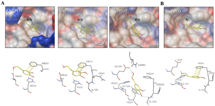
The phenolic compounds containing hydroxytyrosol are the minor components of virgin olive oil (VOO) with the greatest impact on its functional properties and health benefits. Olive breeding for improving the phenolic composition of VOO is strongly dependent on the identification of the key genes determining the biosynthesis of these compounds in the olive fruit and also their transformation during the oil extraction process. In this work, olive polyphenol oxidase (PPO) genes have been identified and fully characterized in order to evaluate their specific role in the metabolism of hydroxytyrosol-derived compounds by combining gene expression analysis and metabolomics data. Four PPO genes have been identified, synthesized, cloned and expressed in Escherichia coli, and the functional identity of the recombinant proteins has been verified using olive phenolic substrates. Among the characterized genes, two stand out: (i) OePPO2 with its diphenolase activity, which is very active in the oxidative degradation of phenols during oil extraction and also seems to be highly involved in the natural defense mechanism in response to biotic stress, and (ii) OePPO3, which codes for a tyrosinase protein, having diphenolase but also monophenolase activity, which catalyzes the hydroxylation of tyrosol to form hydroxytyrosol.

Keywords: Olea europaea L.; hydroxytyrosol; phenolic metabolism; polyphenol oxidase; tyrosinase.


Conflict of interest statement

The authors declare no conflict of interest.

Figures

Figure 1
Figure 1
Multiple sequence alignment of the seven olive PPO proteins identified by transcriptomic analysis. Identical and similar residues are shaded in black and grey, respectively. N-terminal chloroplast transit peptide is squared in blue, and the CuA and CuB domains (tyrosinase domain) are squared in red. The DWL and KFDV domains (middle and C-terminal domains, respectively) are squared in orange, showing the corresponding conserved motif (black line) and the amino acids framing the putative proteolytic region of activation (red line). Strictly conserved amino acids are marked with an asterisk: the copper coordinating histidines (red) of the dicopper centre (CuA and CuB), the cysteines (blue) involved in the disulfide bonds and the thioether bridge and the conserved glutamic acid (water-keeper) and phenylalanine (gatekeeper) residues (black). The two amino acids next to the first and second conserved histidine of CuB (activity controllers) are indicated with a blue triangle.
Figure 2
Figure 2
Phylogenetic tree illustrating relatedness of the seven olive PPO genes identified by transcriptomic analysis to other plant PPO genes. Phylogenetic tree was performed using the neighbor-joining method from MEGA7. Accession numbers are given in Table S2. Red-squared: olive PPOs; solid red line for selected proteins for further studies in this work. Yellow-squared: PPO proteins with diphenolase activity only, as described in the literature. Blue-squared: PPO proteins with both mono- and diphenolase activities as described in the literature.
Figure 3
Figure 3
Heterologous expression and purification of the OePPO proteins. SDS-PAGE followed by a Coomassie blue staining showing the recombinant OePPO purification yield by sepharose-GSH beads affinity chromatography. The results of the inductions for each GST-OePPO recombinant protein (~80 kDa) are shown in lanes T (E. coli total fraction) and S (E. coli soluble fraction). The red arrow shows the corresponding OePPO purified (P) proteins (~55 kDa) from the soluble fraction after removing the GST fusion protein. BL21 refers to control protein extract from untransformed E. coli cells. NR: non-retained fraction; W: wash fraction (1–4 column volumes).
Figure 4
Figure 4
Conversion of tyrosol (Ty) to hydroxytyrosol (HTy) and the hydroxytyrosol quinone (HTy-quinone) by the recombinant OePPO3 protein.
Figure 5
Figure 5
Multiple amino acids alignment of plant PPO regions containing the two activity controller residues, HB1 + 1 and HB2 + 1. Identical and similar residues are shaded in black and grey, respectively. Residues from PPOs with diphenolase activity only have a yellow background, and those from PPOs with di- and monophenolase activities are in green. GenBank Acc. Numbers for plant PPOs are described in Table S3.
Figure 6
Figure 6
Molecular 3D structure of olive PPOs. (A) Coulombic surface coloring for olive PPOs’ molecular 3D structures and their references for modelling. Positively charged amino acid residuals are shaded blue, and negatively charged ones are in red. Activity pocket of the olive PPOs and the proteins used for their molecular modelling are in yellow squares. (B) Active centers of the olive PPOs. Highlighted amino acids in all the enzymes: the six conserved histidines coordinating CuA and CuB (gold spheres) are highlighted as blue sticks; the gatekeeper (phenylalanine) and waterkeeper (glutamic acid) residues are highlighted as red sticks; the first activity controller (HB1 + 1) is shown in green, and the second activity controller (HB2 + 1) is displayed in orange. OePPO1 and OePPO2 are shown as homology models to the CgAUS1 (PDB ID:4z11), and OePPO3 and OePPO4 are shown as homology models to JrPPO1 (PDB ID:5ce9).
Figure 7
Figure 7
Docking analysis of olive PPOs. (A) Potential docking of the diphenolic substrate hydroxytyrosol (HTy) into the active site of OePPO1, OePPO2 and OePPO3 proteins. (B) Potential docking of the monophenolic substrate tyrosol (Ty) into the active site of the OePPO3 enzyme. Surface views of the active pockets are coloured by polarity, with red for negative and blue for positive charged amino acid residues. Amino acids involved in the interaction with the substrate are shown under each docking pose. Hydrogen bonds are shown as green dashed lines, and π-π interactions between aromatic rings are shown as yellow lines.
Figure 8
Figure 8
Relative expression levels of olive OePPO1, OePPO2 and OePPO3 genes in the mesocarp tissue of ‘Picual’, ‘Menya’, ‘Fishomi’, ‘Klon’, ‘Abou Kanani’, and ‘Dokkar’ fruits harvested at the usual ripening stage used for olive oil extraction (stage III).
Figure 9
Figure 9
Relative expression of olive PPO genes by RT-qPCR in cultivar ‘Picual’ fruits under different stress conditions (see Section 3). CON-fly: control sample in fly experiment; fly: fly-attacked fruit; CON-MJ: control sample in methyl jasmonate-treated fruits; MJ: methyl jasmonate-treated fruits. Asterisks (*) mean gene expression values are statistically different (p < 0.05) from their corresponding control.

References

    1. Gouvinhas I., Machado N., Sobreira C., Domínguez-Perles R., Gomes S., Rosa E., Barros A. Critical review on the significance of olive phytochemicals in plant physiology and human health. Molecules. 2017;22:1986. doi: 10.3390/molecules22111986. - DOI - PMC - PubMed
    1. Crespo M.C., Tomé-Carneiro J., Dávalos A., Visioli F. Pharma-nutritional properties of olive oil phenols. Transfer of new findings to human nutrition. Foods. 2018;7:90. doi: 10.3390/foods7060090. - DOI - PMC - PubMed
    1. Parra-Pérez A.M., Pérez-Jiménez A., Gris-Cárdenas I., Bonel-Pérez G.C., Carrasco-Díaz L.M., Mokhtari K., García-Salguero L., Lupiáñez J.A., Rufino-Palomares E.E. Involvement of the PI3K/AKT intracellular signaling pathway in the anticancer activity of hydroxytyrosol, a polyphenol from Olea europaea, in hematological cells and implication of HSP60 levels in its anti-inflamatory activity. Int. J. Mol. Sci. 2022;23:7053. doi: 10.3390/ijms23137053. - DOI - PMC - PubMed
    1. Laghezza Masci V., Bernini R., Villanova N., Clemente M., Cicaloni V., Tinti L., Salvini L., Taddei A.R., Tiezzi A., Ovidi E. In vitro anti-poliferative and apoptotic effects of hydroxytyrosyl oleate on SH-SY5Y human neuroblastoma cells. Int. J. Mol. Sci. 2022;23:12348. doi: 10.3390/ijms232012348. - DOI - PMC - PubMed
    1. European Commission Commission Regulation (EU) No 1018/2013 amending Regulation (EU) No 432/2012 establishing a list of permitted health claims made on foods other than those referring to the reduction of disease risk and to children’s development and heal. Off. J. Eur. Union. 2012;282:43–45.

LinkOut - more resources