

Nanoscale Iron-Based Metal-Organic Frameworks: Incorporation of Functionalized Drugs and Degradation in Biological Media
- PMID: 36834775
- PMCID: PMC9965190
- DOI: 10.3390/ijms24043362
Nanoscale Iron-Based Metal-Organic Frameworks: Incorporation of Functionalized Drugs and Degradation in Biological Media
Abstract
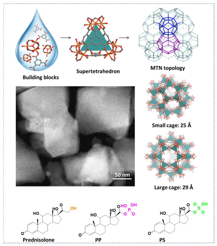
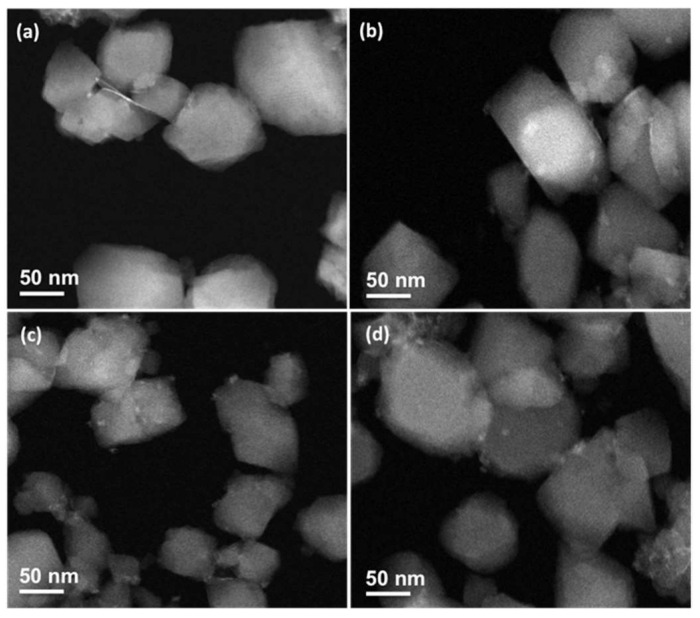
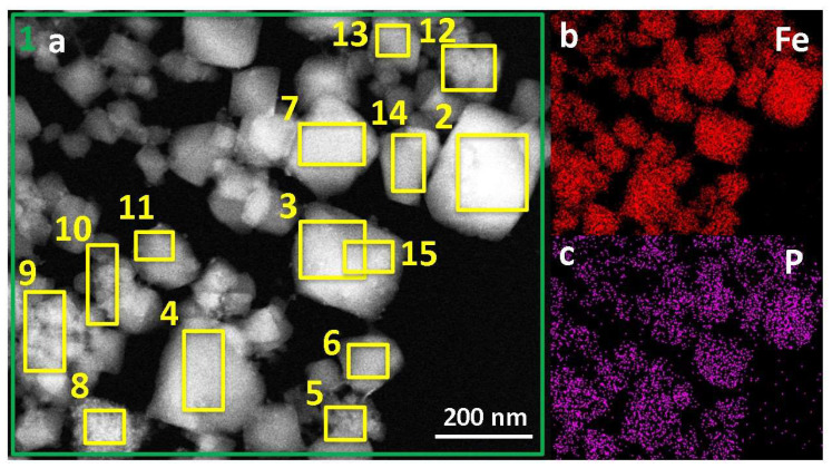
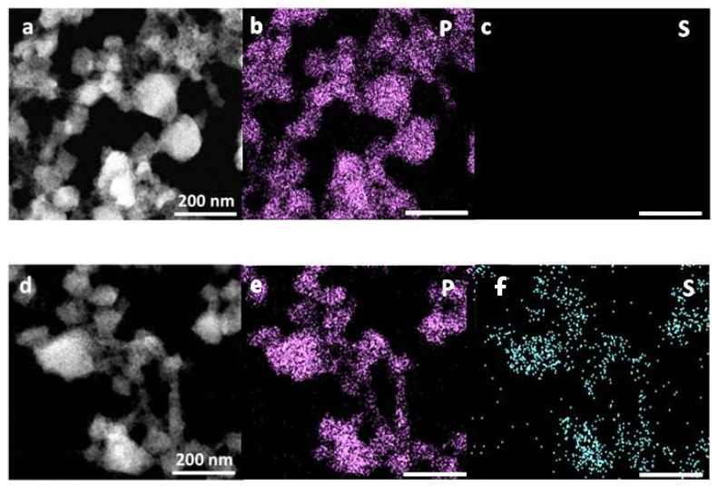
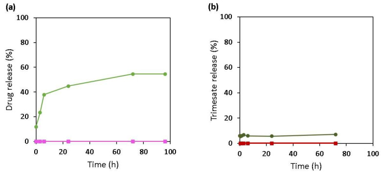
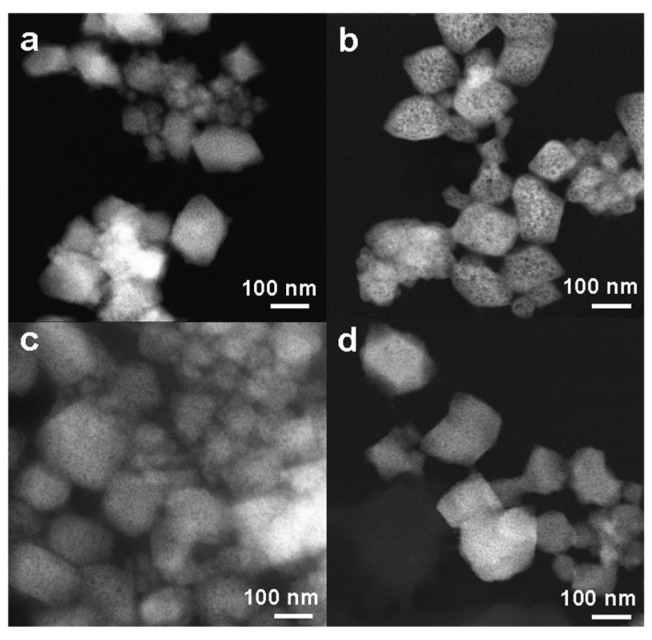
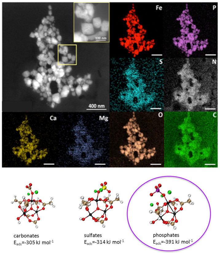
Metal-organic frameworks (MOFs) attract growing interest in biomedical applications. Among thousands of MOF structures, the mesoporous iron(III) carboxylate MIL-100(Fe) (MIL stands for the Materials of Lavoisier Institute) is among the most studied MOF nanocarrier, owing to its high porosity, biodegradability, and lack of toxicity. Nanosized MIL-100(Fe) particles (nanoMOFs) readily coordinate with drugs leading to unprecedented payloads and controlled release. Here, we show how the functional groups of the challenging anticancer drug prednisolone influence their interactions with the nanoMOFs and their release in various media. Molecular modeling enabled predicting the strength of interactions between prednisolone-bearing or not phosphate or sulfate moieties (PP and PS, respectively) and the oxo-trimer of MIL-100(Fe) as well as understanding the pore filling of MIL-100(Fe). Noticeably, PP showed the strongest interactions (drug loading up to 30 wt %, encapsulation efficiency > 98%) and slowed down the nanoMOFs' degradation in simulated body fluid. This drug was shown to bind to the iron Lewis acid sites and was not displaced by other ions in the suspension media. On the contrary, PS was entrapped with lower efficiencies and was easily displaced by phosphates in the release media. Noticeably, the nanoMOFs maintained their size and faceted structures after drug loading and even after degradation in blood or serum after losing almost the totality of the constitutive trimesate ligands. Scanning electron microscopy with high annular dark field (STEM-HAADF) in conjunction with X-Ray energy-dispersive spectrometry (XEDS) was a powerful tool enabling the unraveling of the main elements to gain insights on the MOF structural evolution after drug loading and/or upon degradation.
Keywords: STEM-HAADF; biodegradable nanoparticle; biological media; drug loading; metal–organic frameworks; prednisolone; stability.
Conflict of interest statement
The authors declare no conflict of interest.
Figures
Similar articles
-
Advanced Characterization Methodology to Unravel the Biodegradability of Metal-Organic Framework Nanoparticles in Extremely Diluted Conditions.ACS Appl Mater Interfaces. 2024 Mar 20;16(11):14296-14307. doi: 10.1021/acsami.3c18958. Epub 2024 Mar 7. ACS Appl Mater Interfaces. 2024. PMID: 38452344
-
Biocompatible Fe-Based Micropore Metal-Organic Frameworks as Sustained-Release Anticancer Drug Carriers.Molecules. 2018 Sep 28;23(10):2490. doi: 10.3390/molecules23102490. Molecules. 2018. PMID: 30274195 Free PMC article.
-
Biocompatible porous metal-organic framework nanoparticles based on Fe or Zr for gentamicin vectorization.Eur J Pharm Biopharm. 2018 Nov;132:11-18. doi: 10.1016/j.ejpb.2018.08.013. Epub 2018 Sep 1. Eur J Pharm Biopharm. 2018. PMID: 30179739
-
Recent Advances in Fe-MOF Compositions for Biomedical Applications.Curr Med Chem. 2021;28(30):6179-6198. doi: 10.2174/0929867328666210511014129. Curr Med Chem. 2021. PMID: 33992053 Review.
-
Recent Advances in Metal-Organic Frameworks as Anticancer Drug Delivery Systems: A Review.Anticancer Agents Med Chem. 2021;21(18):2487-2504. doi: 10.2174/1871520621666210119093844. Anticancer Agents Med Chem. 2021. PMID: 33463479 Review.
Cited by
-
Dual-Functioning Metal-Organic Frameworks: Methotrexate-Loaded Gadolinium MOFs as Drug Carriers and Radiosensitizers.Chemistry. 2025 Apr;31(24):e202404106. doi: 10.1002/chem.202404106. Epub 2025 Mar 31. Chemistry. 2025. PMID: 40079794 Free PMC article.
-
MOF-Enhanced Phototherapeutic Wound Dressings Against Drug-Resistant Bacteria.Adv Healthc Mater. 2025 Jan;14(1):e2402418. doi: 10.1002/adhm.202402418. Epub 2024 Oct 26. Adv Healthc Mater. 2025. PMID: 39460484 Free PMC article.
-
Nanoscale coordination polymer Fe-DMY downregulating Poldip2-Nox4-H2O2 pathway and alleviating diabetic retinopathy.J Pharm Anal. 2023 Nov;13(11):1326-1345. doi: 10.1016/j.jpha.2023.05.002. Epub 2023 May 12. J Pharm Anal. 2023. PMID: 38174114 Free PMC article.
-
Iron-MOFs for Biomedical Applications.Adv Healthc Mater. 2025 Mar;14(8):e2402630. doi: 10.1002/adhm.202402630. Epub 2024 Oct 10. Adv Healthc Mater. 2025. PMID: 39388416 Free PMC article. Review.
-
ZnO-Doped Metal-Organic Frameworks Nanoparticles: Antibacterial Activity and Mechanisms.Int J Mol Sci. 2023 Jul 31;24(15):12238. doi: 10.3390/ijms241512238. Int J Mol Sci. 2023. PMID: 37569611 Free PMC article.
References
-
- Horcajada P., Chalati T., Serre C., Gillet B., Sebrie C., Baati T., Eubank J.F., Heurtaux D., Clayette P., Kreuz C., et al. Porous Metal-Organic-Framework Nanoscale Carriers as a Potential Platform for Drug Deliveryand Imaging. Nat. Mater. 2010;9:172–178. - PubMed
-
- Li S., Tan L., Meng X. Nanoscale Metal-Organic Frameworks: Synthesis, Biocompatibility, Imaging Applications, and Thermal and Dynamic Therapy of Tumors. Adv. Funct. Mater. 2020;30:1908924. doi: 10.1002/adfm.201908924. - DOI
-
- Rodriguez-Ruiz V., Maksimenko A., Anand R., Monti S., Agostoni V., Couvreur P., Lampropoulou M., Yannakopoulou K., Gref R. Efficient “Green” Encapsulation of a Highly Hydrophilic Anticancer Drug in Metal-Organic Framework Nanoparticles. J. Drug Target. 2015;23:759–767. - PubMed
MeSH terms
Substances
Grants and funding
LinkOut - more resources
Full Text Sources
Miscellaneous
