Review
. 2023 Feb 8;24(4):3446.
doi: 10.3390/ijms24043446.

Lanthanides-Substituted Hydroxyapatite for Biomedical Applications

Affiliations
Review

Lanthanides-Substituted Hydroxyapatite for Biomedical Applications

María Del Carmen De Lama-Odría et al. Int J Mol Sci. .

Abstract

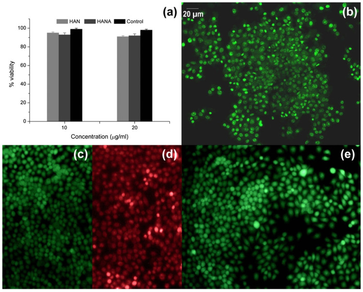
Lately, there has been an increasing demand for materials that could improve tissue regenerative therapies and provide antimicrobial effects. Similarly, there is a growing need to develop or modify biomaterials for the diagnosis and treatment of different pathologies. In this scenario, hydroxyapatite (HAp) appears as a bioceramic with extended functionalities. Nevertheless, there are certain disadvantages related to the mechanical properties and lack of antimicrobial capacity. To circumvent them, the doping of HAp with a variety of cationic ions is emerging as a good alterative due to the different biological roles of each ion. Among many elements, lanthanides are understudied despite their great potential in the biomedical field. For this reason, the present review focuses on the biological benefits of lanthanides and how their incorporation into HAp can alter its morphology and physical properties. A comprehensive section of the applications of lanthanides-substituted HAp nanoparticles (HAp NPs) is presented to unveil the potential biomedical uses of these systems. Finally, the need to study the tolerable and non-toxic percentages of substitution with these elements is highlighted.

Keywords: biolabeling; biomedicine; biosensors; bone regeneration; cancer treatment; cationic ions; cell imaging; doped HAp; hydroxyapatite; implants; lanthanides-substitutions; theragnostics.


Conflict of interest statement

The authors declare no conflict of interest.

Figures

Figure 1
Figure 1
(a) XRD patterns of La-HAp with different La3+ content. In (b), a shift to the left of the XRD peaks can be observed with increasing La3+ concentrations, and in (c), the FTIR profile of the La-HAp powders with different La3+ content is depicted. Reproduced from [36] with permission from Elsevier. Copyright © 2023.
Figure 2
Figure 2
XRD patterns of the La-HAp with different La3+ content and prepared by a high-temperature solid-state reaction process. Peaks attributed to oxyapatite can be observed and a decrease of the diffraction planes of (112) and (300) planes is detectable with higher La3+ concentrations. Reproduced from [37] with permission from Elsevier. Copyright © 2023.
Figure 3
Figure 3
The Wnt signaling pathway. (A) Canonical Wnt signaling cascade. (B) Non-canonical Wnt signaling cascade. APC, adenomatous polyposis coli; CaMKII, calcium/calmodulin-dependent protein kinase type II; CK1α, caseine kinase 1-α; CREB, cyclic AMP-responsive element-binding protein; DAG, diacylglycerol; Dkk, Dickkopf; DSH, disheveled; GSK3β, glycogen synthase kinase-3 β; IP3, inositol 1,4,5-triphosphate; LRP, low-density lipoprotein receptor-related protein; NFAT, nuclear factor of activated T cells; NFκB, nuclear factor κB; PIP2, phosphatidylinositol 4,5-bisphosphate; PKC, protein kinase C; PLC, phospholipase C; sFRPs, secreted frizzled-related proteins; SOST, sclerostin; WIF, Wnt inhibitory factor. For more details on a molecular basis, articles [86,87] can be consulted. Reproduced with permission from [86].
Figure 4
Figure 4
Association of angiogenesis and osteogenesis. (A) VEGF maintains bone homeostasis at physiological levels. Lower levels interrupt osteoblast differentiation and increase osteoclast recruitment. (B) Osteoblasts produce VEGF during bone repair, promoting migration and proliferation of endothelial cells. Endothelial cells later secrete BMP-2, supporting osteoblast differentiation. (C) VEGF regulates Sema3A expression, which suppresses osteoclast differentiation. (D) Sema3A is also responsible for the recruitment of neuropilin 1-expressing (Nrp1+) monocytes. Reproduced with permission from [89].
Figure 5
Figure 5
(a) ALP staining images and (b) alizarin red staining images of the rBMSCs co-cultured with the GdPO4/CTS and β-TCP/CTS scaffolds at days 7 and 14. Reproduced from [124], with permission from Elsevier. Copyright © 2023.
Figure 6
Figure 6
Uptake of HAp, La-HAP and Gd-HAP (the NPs were aforementioned as HAp, La-HAp and Gd-HAp, respectively) into BMSCs and F-actin morphology in the cells upon exposure to the NPs. (A) TEM micrographs of subcellular distribution of HAP (A1,A2), La-HAP (A3,A4) and Gd-HAP after 24 h. Overall cell morphology (left panel), scale bars: 1 μm. Higher magnification of cells in red-boxed areas (right panel, like A2 and A4); scale bars: 200 nm. (B) TEM micrographs of NP adhesion to the cell membrane following incubation with the NPs for 24 h; scale bars: 500 nm. (C) Confocal images of BSMCs showing the F-actin morphology in normal control cells and following incubation with the NPs for 24 h (Rhodamine-phalloidin stained actin filaments). Reproduced from [123] with permission from Elsevier. Copyright © 2023.
Figure 7
Figure 7
Colony plots of different samples after co-culturing with E. coli for 2 h. (a) HA13, (b) 1La-HA13, (c) 1La-HA13/PDA before irradiation, and (d) 1La-HA13/PDA after irradiation. NPs are referred in the review as HAp, 1La-HAp and 1La-HAp/PDA, respectively. Reproduced from [36].
Figure 8
Figure 8
Actin cytoskeleton organization of gingival fibroblasts after 24 h of incubation with uncoated Si substrates and different dextran-coated cerium-doped hydroxyapatite coatings (5CeHAp-D and 10CeHAp-D). F-actin (green) was labeled with phalloidin-phalloidin-fluorescein isothiocyanate (FITC) and nuclei (blue) were counterstained with 4′,6-diamidino-2-phenylindole dihydrochloride (DAPI). Scale bar: 20 µm. Reproduced with permission from [141].
Figure 9
Figure 9
Adhesion of MC3T3-E1 cells on Sm/Gd-HAP coating after culture of (a,b) 5 days and (c,d) 7 days. (Green features correspond to vinculin in the focal adhesion complex.) Reprinted with permission from [143]. Copyright © 2023 American Chemical Society.
Figure 10
Figure 10
(a) MTT assay results. Live/dead image of cells treated with (b) Nd-HAp, (c) negative control, (d) positive control and (e) HAp NPs. Reproduced from [72] with permission from Elsevier. Copyright © 2023.
Figure 11
Figure 11
(a) r1 of DOX/Gd-HAp (indicated as NHA:Gd-DOX in the plot) and Gd-DTPA. Inset: T1 map MRI of different concentrations of NPs and Gd-DTPA. (b) MRI (above) and the color-mapped images (below) of mice post-injection and 15 min after injection of NPs at 10 mg/kg. Scale bar: 5 mm. Reprinted with permission from [73]. Copyright © 2023 American Chemical Society.
Figure 12
Figure 12
Synthesis and application process of the Apt-TDNs-Gd-HAp probe. Reprinted from [151] with the permission of Elsevier. Copyright © 2023.

References

    1. Tite T., Popa A.C., Balescu L.M., Bogdan I.M., Pasuk I., Ferreira J.M.F., Stan G.E. Cationic Substitutions in Hydroxyapatite: Current Status of the Derived Biofunctional Effects and Their In Vitro Interrogation Methods. Materials. 2018;24:2081. - PMC - PubMed
    1. Bose S., Fielding G., Tarafder S., Bandyopadhyay A. Understanding of dopant-induced osteogenesis and angiogenesis in calcium phosphate ceramics. Trends Biotechnol. 2013;31:594–605. - PMC - PubMed
    1. Iconaru S.L., Predoi D., Ciobanu C.S., Motelica-Heino M., Guegan R., Bleotu C. Development of Silver Doped Hydroxyapatite Thin Films for Biomedical Applications. Coatings. 2022;12:341. doi: 10.3390/coatings12030341. - DOI
    1. Iqbal N., Abdul Kadir M.R., Nik Malek N.A.N., Humaimi Mahmood N., Raman Murali M., Kamarul T. Rapid microwave assisted synthesis and characterization of nanosized silver-doped hydroxyapatite with antibacterial properties. Mater. Lett. 2012;89:118–122. doi: 10.1016/j.matlet.2012.08.057. - DOI
    1. Ressler A., Žužić A., Ivanišević I., Kamboj N., Ivanković H. Ionic substituted hydroxyapatite for bone regeneration applications: A review. Open Ceram. 2021;6:100122. doi: 10.1016/j.oceram.2021.100122. - DOI

LinkOut - more resources