Review
. 2023 Feb 11;24(4):3665.
doi: 10.3390/ijms24043665.

Autophagy and the Insulin-like Growth Factor (IGF) System in Colonic Cells: Implications for Colorectal Neoplasia

Affiliations
Review

Autophagy and the Insulin-like Growth Factor (IGF) System in Colonic Cells: Implications for Colorectal Neoplasia

Aldona Kasprzak. Int J Mol Sci. .

Abstract

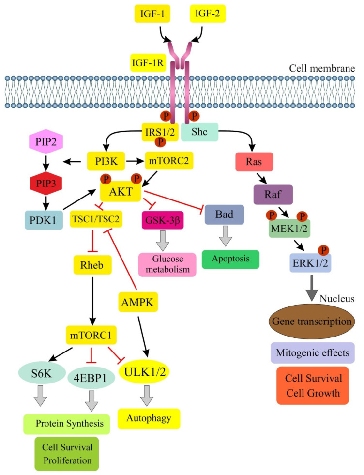
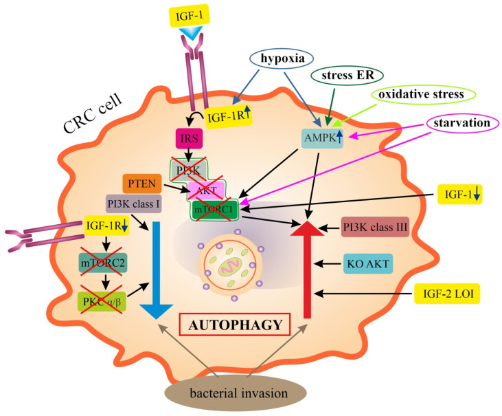
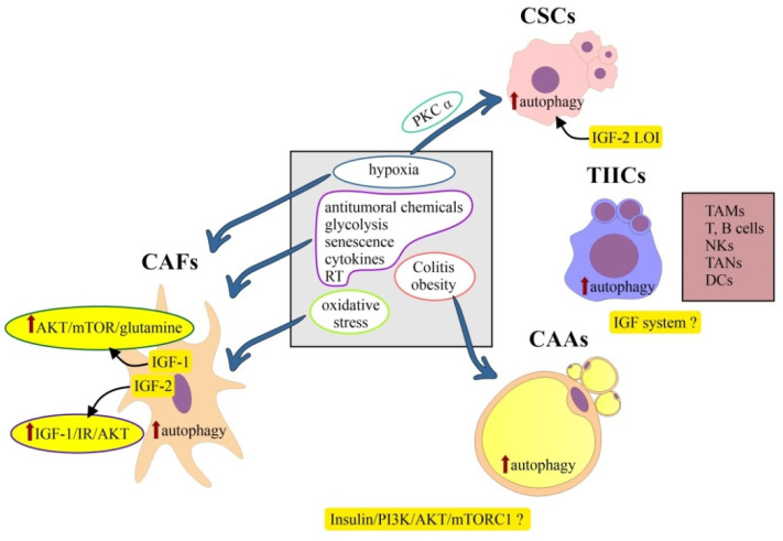
Colorectal cancer (CRC) is one of the most common human malignancies worldwide. Along with apoptosis and inflammation, autophagy is one of three important mechanisms in CRC. The presence of autophagy/mitophagy in most normal mature intestinal epithelial cells has been confirmed, where it has mainly protective functions against reactive oxygen species (ROS)-induced DNA and protein damage. Autophagy regulates cell proliferation, metabolism, differentiation, secretion of mucins and/or anti-microbial peptides. Abnormal autophagy in intestinal epithelial cells leads to dysbiosis, a decline in local immunity and a decrease in cell secretory function. The insulin-like growth factor (IGF) signaling pathway plays an important role in colorectal carcinogenesis. This is evidenced by the biological activities of IGFs (IGF-1 and IGF-2), IGF-1 receptor type 1 (IGF-1R) and IGF-binding proteins (IGF BPs), which have been reported to regulate cell survival, proliferation, differentiation and apoptosis. Defects in autophagy are found in patients with metabolic syndrome (MetS), inflammatory bowel diseases (IBD) and CRC. In neoplastic cells, the IGF system modulates the autophagy process bidirectionally. In the current era of improving CRC therapies, it seems important to investigate the exact mechanisms not only of apoptosis, but also of autophagy in different populations of tumor microenvironment (TME) cells. The role of the IGF system in autophagy in normal as well as transformed colorectal cells still seems poorly understood. Hence, the aim of the review was to summarize the latest knowledge on the role of the IGF system in the molecular mechanisms of autophagy in the normal colon mucosa and in CRC, taking into account the cellular heterogeneity of the colonic and rectal epithelium.

Keywords: autophagy; colonic and rectal epithelial cells; colorectal cancer; insulin-like growth factor (IGF) system; tumor microenvironment (TME) cells.


Conflict of interest statement

The author declares no conflict of interest.

Figures

Figure 1
Figure 1
Schematic illustration of IGF/IGF-1R signaling pathway and its main downstream effectors. Stimulation of serine/threonine kinase AKT (named also PKB) activates mTORC1, leading to increase in protein synthesis. The Ras-Raf-ERK1/2 (MAPK) signaling is also activated to enhance mitogenic effects, cell survival and cell growth. IGF-1 binds mainly to IGF-1R, a typical tyrosine kinase membrane receptor and exerts growth-promoting, anti-apoptotic effects. Initial phosphorylation targets for IGF-1R include IRS 1/2, and among the downstream signaling effectors are PI3K, AKT, mTOR, S6K and Ras-ERK 1/2 (MAPK) pathway components. PI3K kinase, which has two subunits, p85 and p110, in turn phosphorylates a lipid protein, PIP2 to PIP3. PIP3 signals’ proteins such as PDK1 activate AKT by acting on its serine and threonine residues. Activated AKT inhibits GSK-3β and TSC1/TSC2 via phosphorylation. Inactive TSC1/TSC2 is unable to bind Rheb, which subsequently enables the activation of mTORC1 at the surface of lysosome, initiating its effect on S6K, 4EBP1 and ULK1/2, which are responsible for protein synthesis, cell survival, proliferation, delayed apoptosis and inhibition of autophagy. A potential stimulator of autophagy is AMPK signaling. The yellow boxes show the key components of IGF/IGF-1R and AMPK signaling in the regulation of autophagy, the details of which are discussed in the text. Legend: →: lead to; Ʇ: inhibits; AKT (PKB): serine/threonine kinase (protein kinase B); AMPK-5′ adenosine-monophosphate-activated protein kinase; Bad: Bcl-2 associated agonist of cell death; 4EBP1: eukaryotic translation initiation factor-binding protein 4E; GSK-3β: glycogen synthase kinase-3β; IGF: insulin-like growth factor; IGF-1R: IGF receptor type 1; IRS1/2: insulin-receptor substrate 1/2; MAPK: mitogen-activated protein kinase; mTORC1/2: mammalian target of rapamycin complex 1/2; PDK1-3: phosphoinositide-dependent protein kinase 1; PI3K: phosphatidylinositol-3-kinase; PIP2: phosphatidylinositol-4, 5-biphosphate; PIP3: phosphatidylinositol-3,4,5-triphosphate; Rheb: Ras homolog enriched in brain; shc: adaptor protein; S6K: S6 kinase; TSC1/2: tuberous sclerosis complex 1/2; ULK1/2: Unc-51-like autophagy activating kinase 1/2.
Figure 2
Figure 2
Schematic summary of the findings on the role of IGF system in the regulation of autophagy in colorectal cancer (CRC) cells. Neoplastic cells show both induction of autophagy (↑) and inhibition of autophagy (↓). Autophagy is promoted by IGF-1R, whose overexpression under hypoxia inhibits PI3K/AKT/mTORC1 signaling. Inhibition of mTORC1 also occurs under the impact of a decrease in IGF-1 and starvation. Autophagy is directly enhanced by class III PI3K, knockout of AKT and IGF-2 LOI. Inhibition of autophagy in CRC cells is mediated by IGF-1R depletion, through inhibition of mTORC2 and PKC α/β, as well as class I PI3K. Bacterial invasions of the colonic mucosa can both stimulate and inhibit autophagy. Legend: ↑/↓: increase, stimulation/decrease, inhibition; →: lead to; AKT: serine/threonine kinase; AMPK: 5′ adenosine-monophosphate-activated protein kinase; CRC: colorectal cancer; IGF-1/2: insulin-like growth factor 1/2; IGF-1R: IGF receptor type 1; IGF-2 LOI-IGF-2 loss of imprinting; IRS: insulin-receptor substrate; ER: endoplasmic reticulum; KO AKT: knockout of AKT gene; mTORC1/2: mammalian target of rapamycin complex 1/2; PI3K class I or III: phosphatidylinositol-3-kinase class I or III; PKC α/β: protein kinase C alpha/beta; PTEN: phosphatase and tensin homolog deleted on chromosome ten.
Figure 3
Figure 3
Simplified overview of the findings on the pro-autophagic factors (central box) and the potential role of insulin/IGF/PI3K/mTOR signaling (yellow boxes) in the regulation of autophagy in tumor microenvironment cells of colorectal cancer. Detailed mechanisms of regulation of autophagy involving the IGF pathway in TIICs and CAAs are the least understood. Role of IGF-1 in CAFs is mainly explained by the mechanisms of increased proliferation, migration and cell survival of cancer cells via irradiated CAFs, which activate AKT/mTOR/glutamine signaling in these cells. Legend: ↑: increase/stimulation; ?: unknown role; →: lead to; AKT: serine/threonine kinase; AMPK: 5′ adenosine-monophosphate-activated protein kinase; CAAs: cancer-associated adipocytes; CAFs: cancer-associated fibroblasts; CSCs: cancer stem cells; DCs: dendritic cells; IGF-1/2: insulin-like growth factor 1/2; IGF-2 LOI-IGF-2 loss of imprinting; IR: insulin receptor; NKs: natural killer cells; mTOR(C1): mammalian target of rapamycin (complex 1); PI3K: phosphatidylinositol-3-kinase; PKC α: protein kinase C alpha; RT: radiotherapy; TAMs: tumor-associated macrophages; TANs: tumor-associated neutrophils; TIICs: tumor-infiltrating immune cells.

References

    1. Sung H., Ferlay J., Siegel R.L., Laversanne M., Soerjomataram I., Jemal A., Bray F. Global Cancer Statistics 2020: GLOBOCAN Estimates of Incidence and Mortality Worldwide for 36 Cancers in 185 Countries. CA Cancer J. Clin. 2021;71:209–249. doi: 10.3322/caac.21660. - DOI - PubMed
    1. Ahmad R., Singh J.K., Wunnava A., Al-Obeed O., Abdulla M., Srivastava S.K. Emerging trends in colorectal cancer: Dysregulated signaling pathways (Review) Int. J. Mol. Med. 2021;47:14. doi: 10.3892/ijmm.2021.4847. - DOI - PMC - PubMed
    1. Stefani C., Miricescu D., Stanescu-Spinu I.-I., Nica R.I., Greabu M., Totan A.R., Jing M. Growth Factors, PI3K/AKT/mTOR and MAPK Signaling Pathways in Colorectal Cancer Pathogenesis: Where Are We Now? Int. J. Mol. Sci. 2021;22:10260. doi: 10.3390/ijms221910260. - DOI - PMC - PubMed
    1. Vigneri P.G., Tirrò E., Pennisi M.S., Massimino M., Stella S., Romano C., Manzella L. The Insulin/IGF System in Colorectal Cancer Development and Resistance to Therapy. Front. Oncol. 2015;5:230. doi: 10.3389/fonc.2015.00230. - DOI - PMC - PubMed
    1. Kasprzak A., Kwasniewski W., Adamek A., Gozdzicka-Jozefiak A. Insulin-like growth factor (IGF) axis in cancerogenesis. Mutat. Res. Rev. Mutat. Res. 2017;772:78–104. doi: 10.1016/j.mrrev.2016.08.007. - DOI - PubMed

Substances

LinkOut - more resources