Review
. 2023 Feb 12;24(4):3681.
doi: 10.3390/ijms24043681.

The Dilemma of HSV-1 Oncolytic Virus Delivery: The Method Choice and Hurdles

Affiliations
Review

The Dilemma of HSV-1 Oncolytic Virus Delivery: The Method Choice and Hurdles

Guijin Tang et al. Int J Mol Sci. .

Abstract

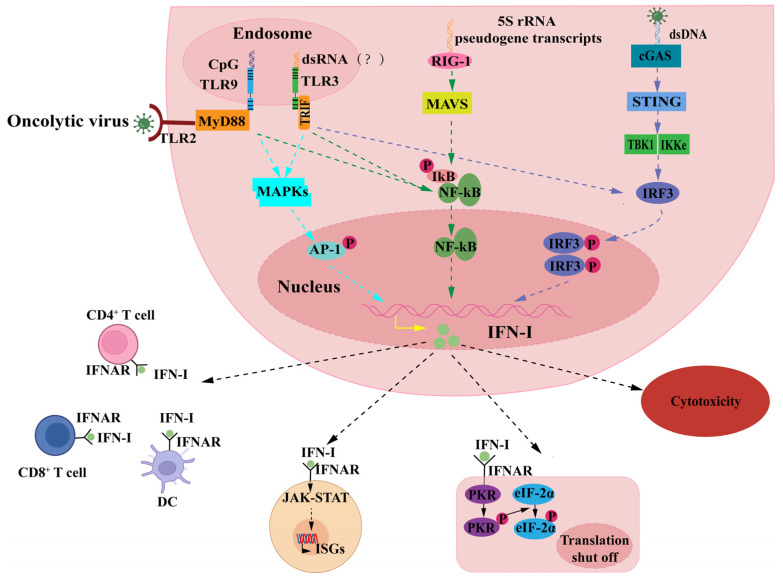
Oncolytic viruses (OVs) have emerged as effective gene therapy and immunotherapy drugs. As an important gene delivery platform, the integration of exogenous genes into OVs has become a novel path for the advancement of OV therapy, while the herpes simplex virus type 1 (HSV-1) is the most commonly used. However, the current mode of administration of HSV-1 oncolytic virus is mainly based on the tumor in situ injection, which limits the application of such OV drugs to a certain extent. Intravenous administration offers a solution to the systemic distribution of OV drugs but is ambiguous in terms of efficacy and safety. The main reason is the synergistic role of innate and adaptive immunity of the immune system in the response against the HSV-1 oncolytic virus, which is rapidly cleared by the body's immune system before it reaches the tumor, a process that is accompanied by side effects. This article reviews different administration methods of HSV-1 oncolytic virus in the process of tumor treatment, especially the research progress in intravenous administration. It also discusses immune constraints and solutions of intravenous administration with the intent to provide new insights into HSV-1 delivery for OV therapy.

Keywords: HSV-1 oncolytic virus therapy; cancer immunotherapy; herpes simplex virus type 1; intravenous injection; mode of administration; oncolytic virus.


Conflict of interest statement

The authors declare no conflict of interest.

Figures

Figure 1
Figure 1
Mechanism of action of oncolytic virus therapy. I: Oncolytic virus (OV) can directly lyse tumor cells and induce immunogenic death of tumor cells, thus, releasing tumor antigens. II and III: Tumor antigens are presented by activated antigen-presenting cells (APCs), which can activate CD8+ T cells in the draining lymph nodes, thus, inducing an organism-specific anti-tumor immune response and playing a role in killing distant metastases. IV: Genetically modified OV promotes antitumor immunity. Genetically engineered OV carrying exogenous genes can improve the immunosuppressed tumor microenvironment (TME) by simultaneously expressing exogenous genes during replication, relieving T-cell suppression, and promoting T-cell activation.
Figure 2
Figure 2
Immune recognition effect induced by intravenous injection of oncolytic virus (OV). When the OV is administered intravenously, the cells of the body’s immune system develop antiviral immunity. Dendritic cells (DC) present viral antigens to T cells and activate them. CD8+ T cells can play a direct toxic role in infected cells by releasing perforin and granzyme and can also cause apoptosis through the Fas pathway; B cells produce neutralizing antibodies by recognizing viral antigens to prevent re-infection of cells, and these antibodies can mediate antibody-dependent cell-mediated cytotoxic effects or activate the complement pathway to produce antiviral effects; macrophages produce large amounts of pro-inflammatory factors during the early stages of infection and phagocytose the virus through cytokinesis; after the downregulation of major histocompatibility complex (MHC) class I molecules on the surface of infected cells, natural killer (NK) cells can recognize the signal to directly kill cells, and infected cells can also activate the receptor through NK cells (AKR activates NK cells to produce pro-inflammatory factors to play a defensive role against viral infections).
Figure 3
Figure 3
Antiviral effect of IFN-I. When viruses infect cells, they can either activate mitogen-activated protein kinase (MAPK) phosphorylation in the nucleus or promote nuclear factor kappa B (NF-κB) dissociation from IκB via myeloid differentiation primary response protein 88 (MyD88) signaling after binding pathogen-associated molecular pattern (PAPMs) to Toll-like receptors 2 (TLR2). Herpes simplex virus type 1 (HSV-1) contains genomes rich in CpG DNA motifs and was demonstrated to activate type I interferon (IFN-I) secretion via TLR9 [75,76]. TLR3 can interact with double-stranded HSV-1, a non-transcribed RNA intermediate, during infection through a MyD88-independent pathway resulting in the activation of NF-κB and interferon regulatory factor (IRF)-3 [77]. In addition, retinoic acid-inducible gene l-like receptors (RIG-I) recognize host 5S rRNA pseudogene transcripts but not HSV-1 genomic-derived RNA after activating IFN-I expression via the NF-κB signaling pathway [78], and viral dsDNA interacts with cyclic GMP-AMP synthase (cGAS) via the cGAS-STING signaling pathway to initiate IFN-I expression. IFN-I binds to IFN receptor and induces more than 200 IFN-stimulated genes (ISGs) via Janus kinase/signal transducers and activators of transcription (JAK-STAT) pathway to produce comprehensive antiviral effects, and it activates relevant immune cells to promote antiviral immunity. This causes cellular transcriptional arrest through the activation of the protein kinase R (PKR) signaling pathway and exerts direct toxic effects on infected cells to resist viral infection.
Figure 4
Figure 4
Strategies to overcome immune clearance and enhance targeting of tumors. These strategies can be applied to prevent the clearance of oncolytic virus (OV) by the body’s antiviral immunity and to improve the targeting of OV when systemic administration of intravenous OV is achieved for the treatment of tumors. (A): Combined with immune inhibitor. (B): Modification of OV using polymers. (C): Use of liposomes to encapsulate OV. (D): Use of nanoparticles to encapsulate OV. (E): Delivery of OV using cell delivery vectors. (F): Genetic editing of OV to improve targeting of tumors.

References

    1. Maurer D.M., Butterfield L.H., Vujanovic L. Melanoma vaccines: Clinical status and immune endpoints. Melanoma Res. 2019;29:109–118. doi: 10.1097/CMR.0000000000000535. - DOI - PMC - PubMed
    1. Forsyth P., Roldan G., George D., Wallace C., Palmer C.A., Morris D., Cairncross G., Matthews M.V., Markert J., Gillespie Y., et al. A phase I trial of intratumoral administration of reovirus in patients with histologically confirmed recurrent malignant gliomas. Mol. Ther. 2008;16:627–632. doi: 10.1038/sj.mt.6300403. - DOI - PubMed
    1. Friedman G.K., Johnston J.M., Bag A.K., Bernstock J.D., Li R., Aban I., Kachurak K., Nan L., Kang K.D., Totsch S., et al. Oncolytic HSV-1 G207 Immunovirotherapy for Pediatric High-Grade Gliomas. N. Engl. J. Med. 2021;384:1613–1622. doi: 10.1056/NEJMoa2024947. - DOI - PMC - PubMed
    1. Patel M.R., Jacobson B.A., Ji Y., Hebbel R.P., Kratzke R.A. Blood Outgrowth Endothelial Cells as a Cellular Carrier for Oncolytic Vesicular Stomatitis Virus Expressing Interferon-beta in Preclinical Models of Non-Small Cell Lung Cancer. Transl. Oncol. 2020;13:100782. doi: 10.1016/j.tranon.2020.100782. - DOI - PMC - PubMed
    1. Monge B M.C., Xie C., Steinberg S.M., Fioraventi S., Walker M., Mabry-Hrones D., Wood B.J., Kleiner D.E., Greten T.F. A phase I/II study of Pexa-Vec oncolytic virus in combination with immune checkpoint inhibition in refractory colorectal cancer. J. Clin. Oncol. 2020;38:117. doi: 10.1200/JCO.2020.38.4_suppl.117. - DOI