

Alzheimer's Disease: An Updated Overview of Its Genetics
- PMID: 36835161
- PMCID: PMC9966419
- DOI: 10.3390/ijms24043754
Alzheimer's Disease: An Updated Overview of Its Genetics
Abstract
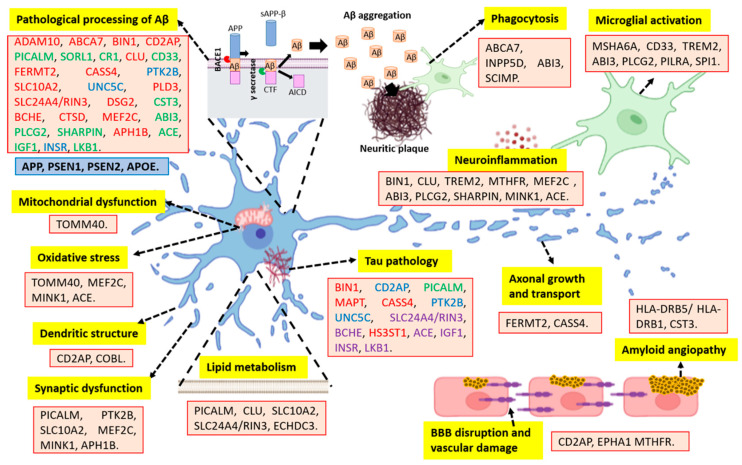
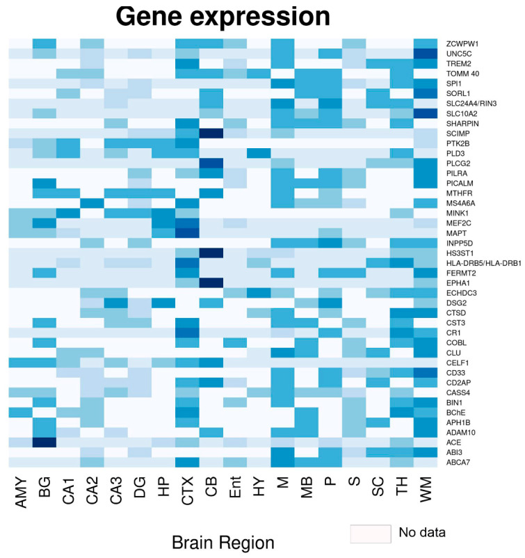
Alzheimer's disease (AD) is the most common neurodegenerative disease in the world. It is classified as familial and sporadic. The dominant familial or autosomal presentation represents 1-5% of the total number of cases. It is categorized as early onset (EOAD; <65 years of age) and presents genetic mutations in presenilin 1 (PSEN1), presenilin 2 (PSEN2), or the Amyloid precursor protein (APP). Sporadic AD represents 95% of the cases and is categorized as late-onset (LOAD), occurring in patients older than 65 years of age. Several risk factors have been identified in sporadic AD; aging is the main one. Nonetheless, multiple genes have been associated with the different neuropathological events involved in LOAD, such as the pathological processing of Amyloid beta (Aβ) peptide and Tau protein, as well as synaptic and mitochondrial dysfunctions, neurovascular alterations, oxidative stress, and neuroinflammation, among others. Interestingly, using genome-wide association study (GWAS) technology, many polymorphisms associated with LOAD have been identified. This review aims to analyze the new genetic findings that are closely related to the pathophysiology of AD. Likewise, it analyzes the multiple mutations identified to date through GWAS that are associated with a high or low risk of developing this neurodegeneration. Understanding genetic variability will allow for the identification of early biomarkers and opportune therapeutic targets for AD.
Keywords: Alzheimer’s disease; GWAS; genetics; loci; molecular mechanisms; neurodegeneration; neuropathology.
Conflict of interest statement
The authors declare no conflict of interest.
Figures
References
-
- 2022 Alzheimer’s disease facts and figures. Alzheimers Dement. 2022;18:700–789. - PubMed
-
- World Health Organization (WHO) [(accessed on 3 November 2022)]. Available online: https://www.who.int/news-room/fact-sheets/detail/dementia.
-
- Matthews K.A., Xu W., Gaglioti A.H., Holt J.B., Croft J.B., Mack D., McGuire L.C. Racial and ethnic estimates of Alzheimer’s disease and related dementias in the United States (2015–2060) in adults aged >/=65 years. Alzheimers Dement. 2019;15:17–24. doi: 10.1016/j.jalz.2018.06.3063. - DOI - PMC - PubMed
Publication types
MeSH terms
Substances
Grants and funding
LinkOut - more resources
Full Text Sources
Medical
