. 2023 Feb 13;24(4):3764.
doi: 10.3390/ijms24043764.

Detection of Breast Cancer-Specific Extracellular Vesicles with Fiber-Optic SPR Biosensor

Affiliations

Detection of Breast Cancer-Specific Extracellular Vesicles with Fiber-Optic SPR Biosensor

Yagmur Yildizhan et al. Int J Mol Sci. .

Abstract

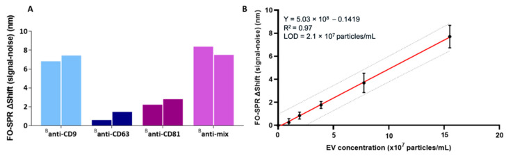
Extracellular vesicles (EVs) have attracted great attention as potential biomarkers for cancer diagnostics. Although several technologies have been developed for EV detection, many of them are still not applicable to clinical settings as they rely on complex EV isolation processes, while lacking sensitivity, specificity or standardization. To solve this problem, we have developed a sensitive breast cancer-specific EV detection bioassay directly in blood plasma using a fiber-optic surface plasmon resonance (FO-SPR) biosensor, previously calibrated with recombinant EVs. First, we established a sandwich bioassay to detect SK-BR-3 EVs by functionalizing the FO-SPR probes with anti-HER2 antibodies. A calibration curve was built using an anti-HER2/Banti-CD9 combination, resulting in an LOD of 2.1 × 10⁷ particles/mL in buffer and 7 × 10⁸ particles/mL in blood plasma. Next, we investigated the potential of the bioassay to detect MCF7 EVs in blood plasma using an anti-EpCAM/Banti-mix combination, obtaining an LOD of 1.1 × 10 8 particles/mL. Finally, the specificity of the bioassay was proven by the absence of signal when testing plasma samples from 10 healthy people unknown to be diagnosed with breast cancer. The remarkable sensitivity and specificity of the developed sandwich bioassay together with the advantages of the standardized FO-SPR biosensor highlight outstanding potential for the future of EV analysis.

Keywords: EpCAM; HER2; biosensors; breast cancer; extracellular vesicles; fiber-optic surface plasmon resonance.


Conflict of interest statement

Jeroen Lammertyn is a board member of FOx Biosystems, a spin-off company of KU Leuven commercializing FO-SPR platforms, next to the principal investigator of the Biosensors group.

Figures

Scheme 1
Scheme 1
Schematic of the different steps from the FO-SPR EV detection sandwich bioassay. The EVs (originating from SK-BR-3 or MCF7 cell lines) are specifically captured by the capture antibodies immobilized on the FO-SPR surface (i.e., anti-HER2 or anti-EpCAM, respectively). Biotinylated detection antibodies (Banti-CD9, Banti-CD63 or Banti-CD81, introduced separately or as a mixture) are used to detect CD9, CD63 and CD81 tetraspanins on the EV surface. Finally, the AuNPs functionalized with anti-biotin antibodies, which recognize multiple biotin labels on the detection antibodies, are used for signal amplification. Antibodies on the FO-SPR surface as well as on the AuNPs have random orientation due to their immobilization through covalent bonds or physical adsorption, respectively. The EVs are depicted in different sizes and colors to simulate the EV heterogeneity in a biological sample. This image was created with Biorender.com (not drawn to scale).
Figure 1
Figure 1
Bar graphs representing the FO-SPR shifts for the immobilization of anti-HER2 antibody using 4 immobilization buffers (10 mM NaAc buffer pH 5.2, 5.4, 5.6, and 50 mM MES buffer pH 6.0) obtained from two independent measurements.
Figure 2
Figure 2
(A) FO-SPR sandwich bioassay established with different antibody combinations for detecting SK-BR-3 EVs in buffer. Bar graphs represent the FO-SPR shifts (obtained after subtracting the negative control, i.e., SPR signal for 0 particles/mL) by combining anti-HER2 as capture antibody with different detection antibodies (Banti-CD9, Banti-CD63, Banti-CD81 or Banti-mix) for detecting SK-BR-3 EVs at 1.55 × 10⁸ particles/mL concentration (measurements shown from two independent experiments). (B) FO-SPR-based detection of a series of SK-BR-3 EV concentrations spiked in the detection buffer (50 mM MES pH 6, 0.01% BSA, 0.01% Tween 20) when using the anti-HER2/Banti-CD9 antibody combination (obtained after subtracting the negative control, i.e., SPR signal for 0 particles/mL). Simple linear regression fitting was performed by GraphPad Prism software (Version 8.0.1, GraphPad Software Inc., MA, USA). The dotted lines indicate the 95% prediction bands for a new observation. Error bars represent one standard deviation (n = 3).
Figure 3
Figure 3
(A) FO-SPR sandwich bioassay with different antibody combinations for detecting SK-BR-3 EVs in 100-fold diluted pooled plasma. Bar graphs represent the FO-SPR shifts from two independent measurements (after subtracting the negative control, i.e., SPR signal for 0 particles/mL) obtained by combining anti-HER2 capture antibody with different detection antibodies (Banti-CD9, Banti-CD63, Banti-CD81 or Banti-mix) for detecting SK-BR-3 EVs at 1.55 × 10⁹ particles/mL concentration. (B) FO-SPR-based detection of a series of SK-BR-3 EV concentrations in 100-fold diluted pooled plasma when using anti-HER2/Banti-CD9 antibody combination (obtained after subtracting the negative control, i.e., SPR signal for 0 particles/mL). Simple linear regression fitting was performed by GraphPad Prism software. The dotted lines indicate the 95% prediction bands for a new observation. Error bars represent one standard deviation (n = 3).
Figure 4
Figure 4
(A) FO-SPR sandwich bioassay with different antibody combinations for detecting MCF7 EVs in 100-fold diluted pooled plasma. Bar graphs represent the FO-SPR shifts (after subtracting the negative control, i.e., SPR signal for 0 particles/mL) obtained from four independent measurements by combining anti-EpCAM capture antibody with different detection antibodies (Banti-CD9, Banti-CD63, Banti-CD81 or Banti-mix) for detecting MCF7 EVs at 1 × 10⁹ particles/mL concentration. (B) FO-SPR-based detection of a series of MCF7 EV concentrations when using anti-EpCAM/Banti-mix antibody combination in 100-fold diluted pooled plasma (obtained after subtracting the negative control, i.e., SPR signal for 0 particles/mL). Simple linear regression fitting was performed by GraphPad Prism software. The dotted lines indicate the 95% prediction bands for a new observation. Error bars represent one standard deviation (n = 3).
Figure 5
Figure 5
Specificity testing of the FO-SPR bioassays in 100-fold diluted plasma using (A) anti-HER2/Banti-CD9 and (B) anti-EpCAM/Banti-mix antibody combinations. P1 to 10 are plasma samples of 10 individual healthy donors unknown to be diagnosed with breast cancer. Control is 100-fold diluted pooled plasma. Two additional controls represent (1) rEVs spiked in 100-fold diluted pooled plasma and (2) SK-BR-3 (in panel (A) or MCF7 EVs (in panel (B) spiked in 100-fold diluted pooled plasma. For panel (A), two independent measurements were performed for all the samples, whereas the number of repetitions was three for panel (B) (although some of the obtained values are close to zero and thus barely visible).

References

    1. Van Niel G., D’Angelo G., Raposo G. Shedding Light on the Cell Biology of Extracellular Vesicles. Nat. Rev. Mol. Cell Biol. 2018;19:213–228. doi: 10.1038/nrm.2017.125. - DOI - PubMed
    1. Ma Y., Dong S., Li X., Kim B.Y.S., Yang Z., Jiang W. Extracellular Vesicles: An Emerging Nanoplatform for Cancer Therapy. Front. Oncol. 2021;10:3340. doi: 10.3389/fonc.2020.606906. - DOI - PMC - PubMed
    1. Xu R., Rai A., Chen M., Suwakulsiri W., Greening D.W., Simpson R.J. Extracellular Vesicles in Cancer—Implications for Future Improvements in Cancer Care. Nat. Rev. Clin. Oncol. 2018;15:617–638. doi: 10.1038/s41571-018-0036-9. - DOI - PubMed
    1. Becker A., Thakur B.K., Weiss J.M., Kim H.S., Peinado H., Lyden D. Extracellular Vesicles in Cancer: Cell-to-Cell Mediators of Metastasis. Cancer Cell. 2016;30:836–848. doi: 10.1016/j.ccell.2016.10.009. - DOI - PMC - PubMed
    1. Simeone P., Bologna G., Lanuti P., Pierdomenico L., Guagnano M.T., Pieragostino D., del Boccio P., Vergara D., Marchisio M., Miscia S., et al. Extracellular Vesicles as Signaling Mediators and Disease Biomarkers across Biological Barriers. Int. J. Mol. Sci. 2020;21:2514. doi: 10.3390/ijms21072514. - DOI - PMC - PubMed