

Use of 3D Spheroid Models for the Assessment of RT Response in Head and Neck Cancer
- PMID: 36835181
- PMCID: PMC9963786
- DOI: 10.3390/ijms24043763
Use of 3D Spheroid Models for the Assessment of RT Response in Head and Neck Cancer
Abstract
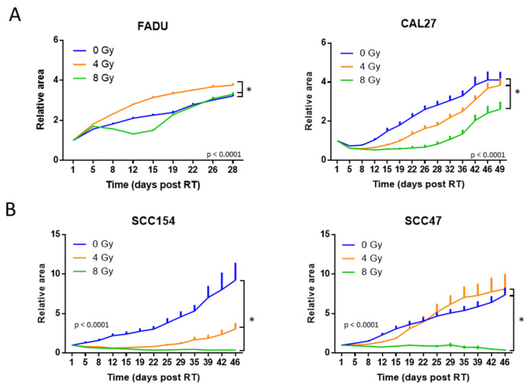
Radiotherapy (RT) is a key player in the treatment of head and neck cancer (HNC). The RT response, however, is variable and influenced by multiple tumoral and tumor microenvironmental factors, such as human papillomavirus (HPV) infections and hypoxia. To investigate the biological mechanisms behind these variable responses, preclinical models are crucial. Up till now, 2D clonogenic and in vivo assays have remained the gold standard, although the popularity of 3D models is rising. In this study, we investigate the use of 3D spheroid models as a preclinical tool for radiobiological research by comparing the RT response of two HPV-positive and two HPV-negative HNC spheroid models to the RT response of their corresponding 2D and in vivo models. We demonstrate that HPV-positive spheroids keep their higher intrinsic radiosensitivity when compared to HPV-negative spheroids. A good correlation is found in the RT response between HPV-positive SCC154 and HPV-negative CAL27 spheroids and their respective xenografts. In addition, 3D spheroids are able to capture the heterogeneity of RT responses within HPV-positive and HPV-negative models. Moreover, we demonstrate the potential use of 3D spheroids in the study of the mechanisms underlying these RT responses in a spatial manner by whole-mount Ki-67 and pimonidazole staining. Overall, our results show that 3D spheroids are a promising model to assess the RT response in HNC.
Keywords: 3D spheroid models; head and neck cancer; human papillomavirus; radiotherapy.
Conflict of interest statement
The authors M.W., R.D. and S.N. declare no conflicts of interest. L.J.D. has shares in the company Convert Pharmaceuticals, and has a non-issue patent on LSRT (PCT/P126537PC00), licensed to Varian. None of the above entities were involved in the preparation of this paper. The funders had no role in the design of the study; in the collection, analyses, or interpretation of the data; in the writing of the manuscript; or in the decision to publish the results.
Figures


Similar articles
-
Effectiveness of PARP inhibition in enhancing the radiosensitivity of 3D spheroids of head and neck squamous cell carcinoma.Front Oncol. 2022 Aug 16;12:940377. doi: 10.3389/fonc.2022.940377. eCollection 2022. Front Oncol. 2022. PMID: 36052247 Free PMC article.
-
Intrinsic Radiosensitivity Is Not the Determining Factor in Treatment Response Differences between HPV Negative and HPV Positive Head and Neck Cancers.Cells. 2020 Jul 27;9(8):1788. doi: 10.3390/cells9081788. Cells. 2020. PMID: 32727072 Free PMC article.
-
Analysis of human leukocyte antigen associations in human papillomavirus-positive and -negative head and neck cancer: Comparison with cervical cancer.Cancer. 2022 May 15;128(10):1937-1947. doi: 10.1002/cncr.34148. Epub 2022 Feb 17. Cancer. 2022. PMID: 35176174 Free PMC article.
-
Enhanced Radiation Sensitivity of Human Papillomavirus-Driven Head and Neck Cancer: Focus on Immunological Aspects.Front Immunol. 2019 Dec 3;10:2831. doi: 10.3389/fimmu.2019.02831. eCollection 2019. Front Immunol. 2019. PMID: 31849993 Free PMC article. Review.
-
Increased radiosensitivity of HPV-positive head and neck cancers: Molecular basis and therapeutic perspectives.Cancer Treat Rev. 2015 Dec;41(10):844-52. doi: 10.1016/j.ctrv.2015.10.001. Epub 2015 Oct 13. Cancer Treat Rev. 2015. PMID: 26476574 Review.
Cited by
-
In Vitro Models of Head and Neck Cancer: From Primitive to Most Advanced.J Pers Med. 2023 Nov 3;13(11):1575. doi: 10.3390/jpm13111575. J Pers Med. 2023. PMID: 38003890 Free PMC article. Review.
-
Evaluating ECM stiffness and liver cancer radiation response via shear-wave elasticity in 3D culture models.Radiat Oncol. 2024 Sep 27;19(1):128. doi: 10.1186/s13014-024-02513-7. Radiat Oncol. 2024. PMID: 39334323 Free PMC article.
-
Radiobiology of Proton Therapy in Human Papillomavirus-Negative and Human Papillomavirus-Positive Head and Neck Cancer Cells.Cancers (Basel). 2024 May 22;16(11):1959. doi: 10.3390/cancers16111959. Cancers (Basel). 2024. PMID: 38893080 Free PMC article.
-
Three-dimensional in vitro models in head and neck cancer: current trends and applications.Med Oncol. 2025 May 5;42(6):194. doi: 10.1007/s12032-025-02737-x. Med Oncol. 2025. PMID: 40320444 Review.
References
-
- Machiels J.P., René Leemans C., Golusinski W., Grau C., Licitra L., Gregoire V. Reprint of “Squamous Cell Carcinoma of the Oral Cavity, Larynx, Oropharynx and Hypopharynx: EHNS-ESMO-ESTRO Clinical Practice Guidelines for Diagnosis, Treatment and Follow-Up”. Oral Oncol. 2020;31:1462–1475. doi: 10.1016/j.oraloncology.2020.105042. - DOI - PubMed
MeSH terms
Grants and funding
- NA/Emmanuel van der Schueren fellowship for pre-doctoral researchers from Kom op Tegen Kanker
- NA/Emmanuel van der Schueren fellowship for postdoctoral researchers from Kom op Tegen Kanker
- 18B4122N/Flemish Foundation of Scientific Research
- 1521019N/Flemish Foundation of Scientific Research
- C24/17/073/KU Leuven Research Fund
LinkOut - more resources
Full Text Sources
Medical
