. 2023 Feb 14;24(4):3794.
doi: 10.3390/ijms24043794.

Vorinostat Improves Myotonic Dystrophy Type 1 Splicing Abnormalities in DM1 Muscle Cell Lines and Skeletal Muscle from a DM1 Mouse Model

Affiliations

Vorinostat Improves Myotonic Dystrophy Type 1 Splicing Abnormalities in DM1 Muscle Cell Lines and Skeletal Muscle from a DM1 Mouse Model

Nafisa Neault et al. Int J Mol Sci. .

Abstract

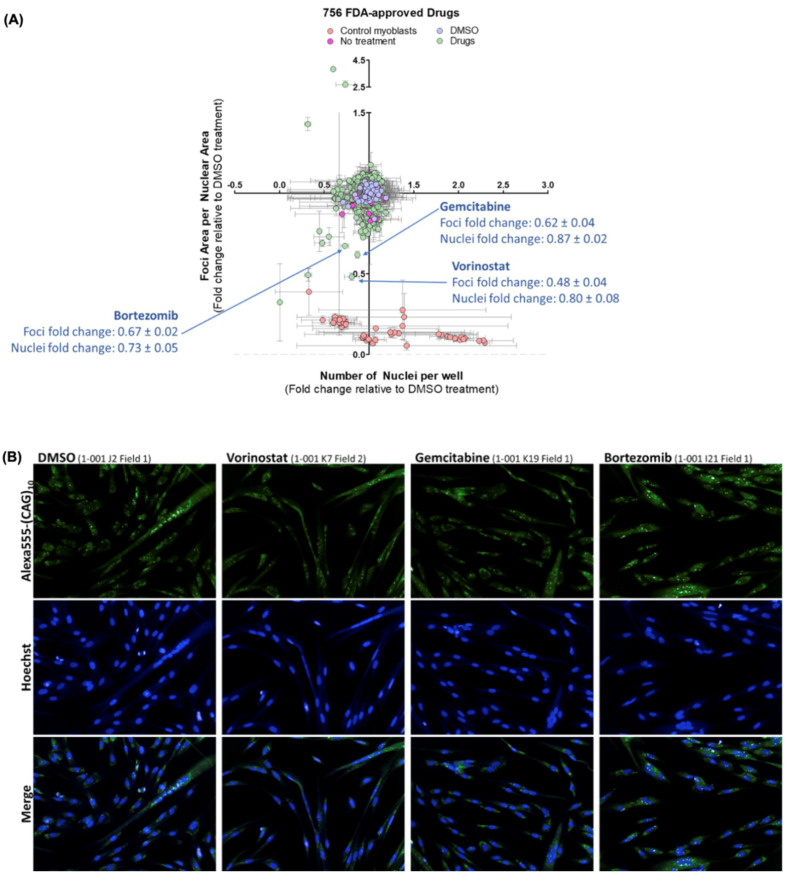
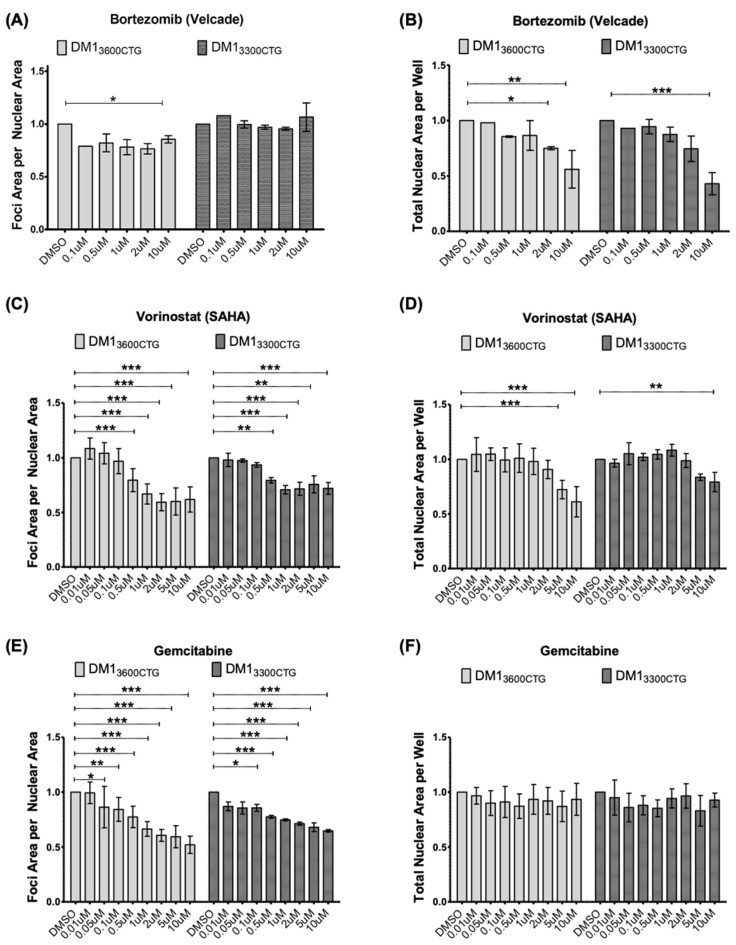
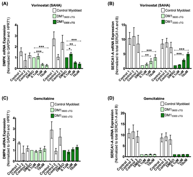
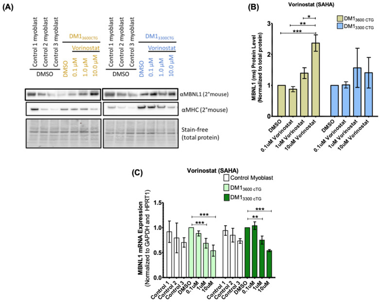
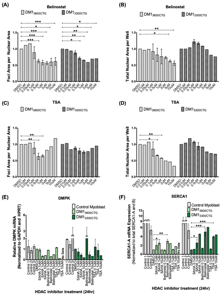
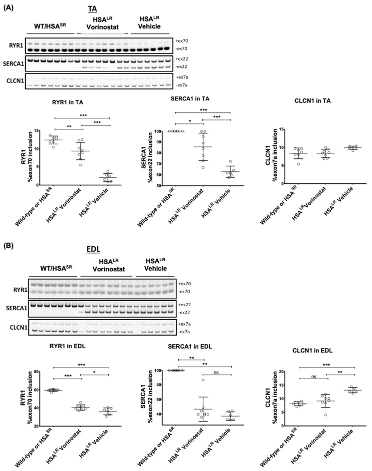
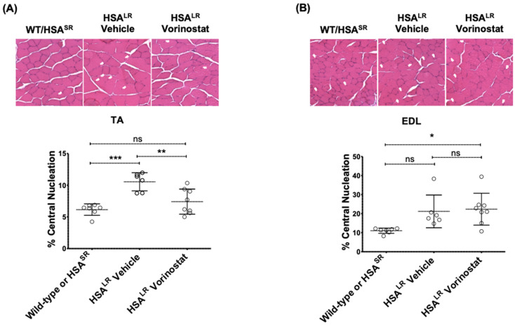
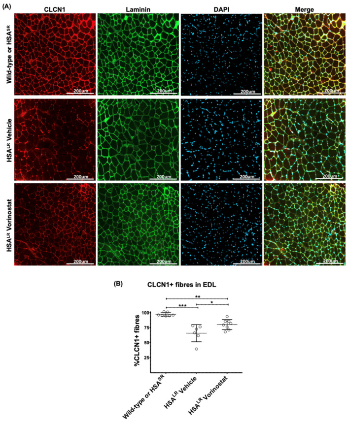
Myotonic dystrophy type 1 (DM1), the most common form of adult muscular dystrophy, is caused by an abnormal expansion of CTG repeats in the 3' untranslated region of the dystrophia myotonica protein kinase (DMPK) gene. The expanded repeats of the DMPK mRNA form hairpin structures in vitro, which cause misregulation and/or sequestration of proteins including the splicing regulator muscleblind-like 1 (MBNL1). In turn, misregulation and sequestration of such proteins result in the aberrant alternative splicing of diverse mRNAs and underlie, at least in part, DM1 pathogenesis. It has been previously shown that disaggregating RNA foci repletes free MBNL1, rescues DM1 spliceopathy, and alleviates associated symptoms such as myotonia. Using an FDA-approved drug library, we have screened for a reduction of CUG foci in patient muscle cells and identified the HDAC inhibitor, vorinostat, as an inhibitor of foci formation; SERCA1 (sarcoplasmic/endoplasmic reticulum Ca2+-ATPase) spliceopathy was also improved by vorinostat treatment. Vorinostat treatment in a mouse model of DM1 (human skeletal actin-long repeat; HSALR) improved several spliceopathies, reduced muscle central nucleation, and restored chloride channel levels at the sarcolemma. Our in vitro and in vivo evidence showing amelioration of several DM1 disease markers marks vorinostat as a promising novel DM1 therapy.

Keywords: myotonic dystrophy type 1; new treatment; rare diseases; vorinostat.


Conflict of interest statement

The authors declare no conflict of interest.

Figures

Figure 1
Figure 1
FDA small-molecule screen identified vorinostat, gemcitabine, and bortezomib as potential down-regulators of CUG foci. DM13600CTG myoblasts were serum-starved in 384-well plates for 7 days and treated with 2 μM of drugs for 24 h. Post-treatment, cells were stained with Hoechst for DNA and Alexa555-(CAG)10 for CUG RNA foci. Foci area per nuclear area and number of nuclei per well were quantified using Columbus and normalized to DMSO control data per plate. (A) Data is presented as the average fold-change relative to DMSO treatment (n = 3, error bars represent SD). (B) Representative images of top hits for small molecules which reduce foci. Full data set is summarized in Supplementary Table S1.
Figure 2
Figure 2
Bortezomib minimally reduced foci; vorinostat (SAHA) and gemcitabine reduced foci in both DM13600CTG and DM13300CTG differentiated myoblasts. DM13600CTG and DM13300CTG myoblasts were serum-starved in 384-well plates for 7 days and treated with 0.1–10 μM of (A,B) bortezomib, (C,D) vorinostat (SAHA), and (E,F) gemcitabine. Post-treatment, cells were fixed with 4% PFA, DNA was stained with Hoechst and CUG RNA foci were probed by Alexa555-(CAG)10 fluorescent oligo. (A,C,E) Foci area per nuclear area and (B,D,F) total nuclear area per well (to assess treatment-associated toxicity) were quantified using Columbus and normalized to DMSO control; data is presented as fold-change relative to DMSO treatment (n ranges from 1 (for Bortezomib 0.1 µM) to 8, two-way ANOVA; error bars represent SD). * p < 0.05, ** p < 0.01, *** p < 0.001.
Figure 3
Figure 3
Vorinostat (SAHA), but not gemcitabine, reduced DMPK mRNA levels and rescued the SERCA1-A splice product in both DM13600CTG and DM13300CTG differentiated myoblasts. Control DM13600CTG and DM13300CTG myoblasts were serum-starved in 6-well plates for 7 days. DM1 cells were treated with DMSO alone or 0.1, 1 or 10 μM of (A,B) vorinostat (SAHA) or (C,D) gemcitabine; control myoblasts were treated with DMSO and used for baseline quantification in unaffected cells. RNA was extracted (RNeasy micro kit, Qiagen) and reverse-transcribed to cDNA (iScript Advanced RT kit, Biorad). RT-qPCR was performed (iQ Sybr green supermix, Biorad) to assess mRNA levels (n = 6, two-way ANOVA; error bars represent SD). * p < 0.05, ** p < 0.01, *** p < 0.001.
Figure 4
Figure 4
Vorinostat increased MBNL1 protein in DM13600CTG differentiated myoblasts but decreased MBNL1 mRNA. (A,B) DM13600CTG and DM13300CTG myoblasts were serum-starved in 6 cm plates for 7 days and treated with DMSO alone or 0.1, 1 and 10 μM of vorinostat (SAHA). 24 h post-treatment, cells were trypsinized and extracted for protein using RIPA lysis buffer for western blot analysis. (A) Western blot images; (B) quantification of MBNL1 protein normalized to total protein and presented as fold-change relative to DMSO control (n = 2; two-way ANOVA). (C) DM13600CTG and DM13300CTG myoblasts were serum-starved in 6-well plates or 6 cm plates and treated with DMSO alone or 0.1, 1 and 10 μM of vorinostat (SAHA). 24 h post-treatment, cells were lysed directly on plate (6-well plate) or trypsinized and the pellet lysed (6 cm plate). RNA was extracted (RNeasy micro kit, Qiagen) and reverse-transcribed to cDNA (iScript Advanced RT kit, Biorad). RT-qPCR was performed (iQ Sybr green supermix, Biorad) to assess mRNA levels (n = 6, two-way ANOVA; error bars represent SD). * p < 0.05, ** p < 0.01, *** p < 0.001.
Figure 5
Figure 5
Two additional pan-HDAC inhibitors, belinostat and trichostatin A (TSA), were validated to reduce foci, decrease DMPK mRNA, and rescue SERCA1 splicing in differentiated DM1 myoblasts. (AD) DM13600CTG and DM13300CTG myoblasts were grown and/or serum-starved in 384-well plates for 7 days and treated with DMSO alone or 0.01, 0.05, 0.1, 0.5, 1, 2, 5 and 10 μM of HDAC inhibitor. 24 h post-treatment, cells were fixed with 4% PFA, DNA was stained with Hoechst and CUG RNA foci were probed by Alexa555-(CAG)10 fluorescent oligo. Foci area per nuclear area and total nuclear area per well (to assess treatment-associated toxicity) were quantified using Columbus and normalized to DMSO control; data is presented as fold-change relative to DMSO treatment (n ranges from 1 to 3, two-way ANOVA; error bars represent SD). (E-F) DM13600CTG and DM13300CTG myoblasts were serum-starved in 6-well plates for 7 days. DM1 cells were treated with DMSO alone or 0.1, 1 or 10 μM of vorinostat (SAHA), belinostat, and TSA. RNA was extracted (RNeasy micro kit, Qiagen) and reverse-transcribed to cDNA (iScript Advanced RT kit, Biorad). RT-qPCR was performed (iQ Sybr green supermix, Biorad) to assess mRNA levels of (E) DMPK and (F) SERCA1 splicing. (n ranges from 1 to 2, two-way ANOVA; error bars represent SD). * p < 0.05, ** p < 0.01, *** p < 0.001.
Figure 6
Figure 6
Vorinostat (SAHA) showed promising therapeutic effects in the DM1 HSALR mouse model, with greater impact in TA than EDL. Mice at approximately 4 weeks of age were injected (IP) daily with vehicle or 50 mg/kg vorinostat (SAHA) for 4 weeks. The mice were sacrificed by lethal injection and cervical dislocation. Skeletal muscle tissue from one hind leg was frozen in OCT for sectioning and imaging, and skeletal muscle tissue from the other hind leg was flash frozen in liquid nitrogen for RNA workup. Analysis of DM1 related spliceopathy. Flash frozen tissue was ground to a powder and a portion used for Trizol RNA extraction using 5 the Purelink RNA mini kit (Invitrogen) and reverse transcribed to cDNA (iScript Advanced RT kit, BioRad). RT-sqPCR products using cDNA from (A) TA and (B) EDL were resolved on 7% acrylamide gel, stained with Gel Red and imaged using the ChemiDoc (BioRad). Quantification of transcript ratios was done using ImageLab (BioRad). (n ranges from 6 to 8, two-way ANOVA; error bars represent SD). * p < 0.05, ** p < 0.01, *** p < 0.001. Abbreviations, EDL, extensor digitorum longus; TA, tibialis anterior.
Figure 7
Figure 7
Vorinostat (SAHA) reduced central nucleation in TA muscle of DM1 HSALR mouse model. Mice at approximately 4 weeks of age were injected (IP) daily with vehicle or 50 mg/kg vorinostat (SAHA) for 4 weeks. The mice were sacrificed by lethal injection and cervical dislocation. Skeletal muscle tissue from one hind leg was frozen in OCT for sectioning and imaging, and central nucleation was assessed in (A) TA and (B) EDL muscle. Tissues flash frozen in OCT were sectioned at 10 μm thickness and subjected to H&E staining. Brightfield images were taken at 20× using EVOS cell imaging system and whole sections were manually counted for central nucleation. (n ranges from 6 to 8, two-way ANOVA; error bars represent SD). * p < 0.05, ** p < 0.01, *** p < 0.001. Abbreviations, EDL, extensor digitorum longus; ns, not significant; TA, tibialis anterior.
Figure 8
Figure 8
Vorinostat (SAHA) restored chloride channel protein levels in EDL muscle of DM1 HSALR mouse model. Mice at approximately 4 weeks of age were injected (IP) daily with vehicle or 50 mg/kg vorinostat (SAHA) for 4 weeks. The mice were sacrificed by lethal injection and cervical dislocation. Skeletal muscle tissue from one hind leg was frozen in OCT for sectioning and imaging. (A) Tissues flash frozen in OCT were sectioned at 10 μm thickness and subjected to immunofluorescence staining using antibodies against chloride channels (CLCN1) and laminin, and counterstained with DAPI. Brightfield images were taken at 20× using EVOS 5 cell imaging system. (B) CLCN1-positive fibers and lamini-positive fibers were manually counted using ImageJ and the percentage of CLCN1-positive fibers was calculated relative to total fibers as represented by lamini-positive fibers. (n ranges from 6 to 8, two-way ANOVA; error bars represent SD). * p < 0.05, ** p < 0.01, *** p < 0.001. Abbreviations, EDL, extensor digitorum longus.

References

    1. Suominen T., Bachinski L.L., Auvinen S., Hackman P., Baggerly K.A., Angelini C., Peltonen L., Krahe R., Udd B. Population frequency of myotonic dystrophy: Higher than expected frequency of myotonic dystrophy type 2 (DM2) mutation in Finland. Eur. J. Hum. Genet. 2011;19:776–782. doi: 10.1038/ejhg.2011.23. - DOI - PMC - PubMed
    1. Johnson N.E., Butterfield R.J., Mayne K., Newcomb T., Imburgia C., Dunn D., Duval B., Feldkamp M.L., Weiss R.B. Population-based prevalence of myotonic dystrophy type 1 using genetic analysis of statewide blood screening program. Neurology. 2021;96:e1045–e1053. - PMC - PubMed
    1. Reardon W., MacMillan J., Myring J., Harley H., Rundle S., Beck L., Harper P., Shaw D. Cataract and myotonic dystrophy: The role of molecular diagnosis. Br. J. Ophthalmol. 1993;77:579–583. doi: 10.1136/bjo.77.9.579. - DOI - PMC - PubMed
    1. Antonini G., Clemenzi A., Bucci E., Morino S., Garibaldi M., Sepe-Monti M., Giubilei F., Novelli G. Erectile dysfunction in myotonic dystrophy type 1 (DM1) J. Neurol. 2009;256:657. doi: 10.1007/s00415-009-0912-4. - DOI - PubMed
    1. Voermans N.C., Erasmus C.E., Ockeloen C.W., Van Engelen B.G., Eggink C.A. Primary cataract as a key to recognition of myotonic dystrophy type 1. Eur. J. Ophthalmol. 2015;25:e46–e49. doi: 10.5301/ejo.5000565. - DOI - PubMed

Substances

LinkOut - more resources