Review
. 2023 Feb 14;24(4):3803.
doi: 10.3390/ijms24043803.

Tuning the Wavelength: Manipulation of Light Signaling to Control Plant Defense

Affiliations
Review

Tuning the Wavelength: Manipulation of Light Signaling to Control Plant Defense

Susan Breen et al. Int J Mol Sci. .

Abstract

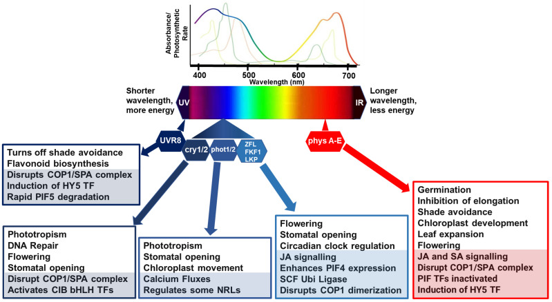
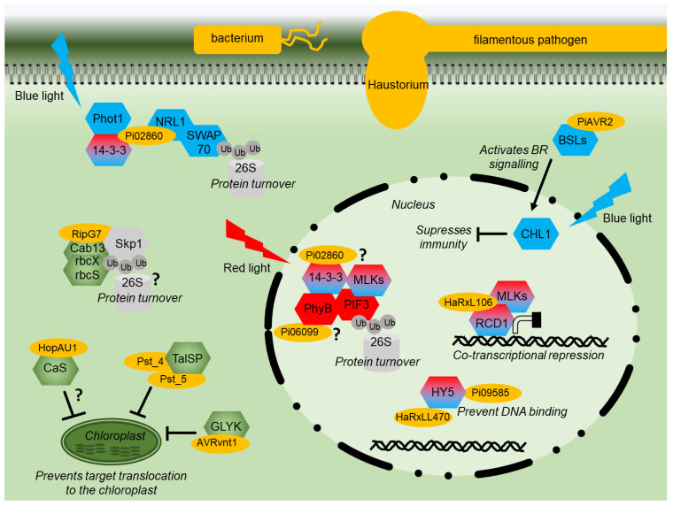
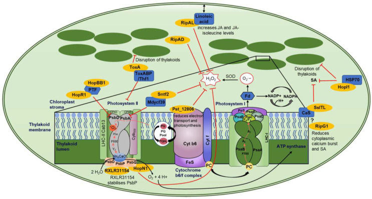
The growth-defense trade-off in plants is a phenomenon whereby plants must balance the allocation of their resources between developmental growth and defense against attack by pests and pathogens. Consequently, there are a series of points where growth signaling can negatively regulate defenses and where defense signaling can inhibit growth. Light perception by various photoreceptors has a major role in the control of growth and thus many points where it can influence defense. Plant pathogens secrete effector proteins to manipulate defense signaling in their hosts. Evidence is emerging that some of these effectors target light signaling pathways. Several effectors from different kingdoms of life have converged on key chloroplast processes to take advantage of regulatory crosstalk. Moreover, plant pathogens also perceive and react to light in complex ways to regulate their own growth, development, and virulence. Recent work has shown that varying light wavelengths may provide a novel way of controlling or preventing disease outbreaks in plants.

Keywords: immunity; light; pathogen effectors; pathogenicity; plant; signaling.


Conflict of interest statement

The authors declare no conflict of interest.

Figures

Figure 1
Figure 1
Simplified model of light perception and associated molecular and physiological processes. Lines, above the light spectrum, represent the light absorption spectra for the major photosynthetic pigments, chlorophyll a (lime green), b (apple green) and carotenoids (brown/green line). The rainbow line highlights photosynthetic rate relative to wavelength. Below the light spectra indicates multiple photoreceptors that have evolved to detect blue and red wavelengths. In general, in poor light quality, stabilization of photomorphogenesis-promoting factors occurs, promoting growth in the direction of light. Photomorphogenic responses are regulated by phytochromes (phyA to E), which absorb RL/FRL; cryptochromes (cry 1/2), which are activated by BL/UV-A; phototropins (phot 1/2), activated by BL/UV-A; and Zeitlupe family members (Ztl, fkf1, lkp2) activated by BL/UV-A and the UV-B photoreceptor (UV resistance locus; UVR8). Light activation of phytochromes, cryptochromes, and UVR8 promotes interactions with transcription factors to relay responses to light conditions. CONSTITUTIVE PHOTOMORPHOGENIC 1 and SUPPRESSOR OF PHYTOCHROME A (COP1/SPA) complex act as substrate adaptors for CULLIN4 (CUL4) E3 ligase complexes, key repressors of light responses in darkness. Under high RL:FRL conditions, ELONGATED HYPOCOTYL 5 (HY5) is induced and Phytochrome-Interacting Factor (PIF) activities are inactivated by phyB to suppress shade avoidance responses such as elongation. Under UV and blue lights, COP1/SPA is disrupted to suppress unnecessary and costly photomorphogenesis responses. UVR8 induces HY5 activity, and the binding of UVR8 to COP1 in UV-B disrupts PIF5 stabilization, rapidly lowering PIF5 abundance in sunlight by degradation via ubiquitination and proteasome-mediated protein degradation. CRYs disrupt the COP1/SPA complex which activates cryptochrome-interacting basic helix–loop–helix (CIB1) and CIB1-related proteins to promote floral initiation, DNA repair, and stomatal opening. Phots have their own kinase activities associated with regulation of physiological responses to optimize photosynthesis, such as stomatal opening to regulate CO2 uptake and water loss, chloroplast movements, calcium fluxes, and leaf positioning. Activated phots interact with some NRL family members as a substrate adaptor c of a CULLIN3 E3 ubiquitin ligase. Zeitlupe family members disrupt COP1 dimerization, enhance PIF4 expression, and regulate responses such as flowering time, JA biosynthesis, and stomatal opening. COP1/SPA can also target some additional E3 ligases or kinases for degradation causing indirect increases and stabilization of downstream transcription factors, such as PIFs. In each box, the lower shaded sections highlight molecular mechanisms, and the non-shaded, upper sections features physiological processes.
Figure 2
Figure 2
Effector modes of action diagram. Diagram of a plant cell showing the modes of action of various pathogen effector proteins (yellow). Signaling components of red or blue light signaling are shown in red or blue, respectively. Shared components are shaded red and blue. Proteins with a chloroplast function are shown in green. Components of the proteasome are indicated in grey. Interactions that remain to be fully characterized are indicated by “?”.
Figure 3
Figure 3
Chloroplast-localized effector proteins and their targets. This figure shows a representative image of a chloroplast and the effector–target interactions with downstream mode of action. The enlarged box shows a schematic of the thylakoid membrane (dark green) with PSII, cytochrome b6/f complex, and PSI (various colors to better visualize subunits) along with effectors targeting various components (dark blue) of these complexes. Effectors are shown in yellow, inhibitory signaling is shown by a red line, while electron transport is shown by black arrows. The ? represents speculative interactions, and double-ended arrows represent movement.

Similar articles

Cited by

References

    1. Ballaré C.L., Mazza C.A., Austin A.T., Pierik R. Canopy Light and Plant Health. Plant Physiol. 2012;160:145–155. doi: 10.1104/pp.112.200733. - DOI - PMC - PubMed
    1. Beattie G.A., Hatfield B.M., Dong H., McGrane R.S. Seeing the Light: The Roles of Red- and Blue-Light Sensing in Plant Microbes. Annu. Rev. Phytopathol. 2018;56:41–66. doi: 10.1146/annurev-phyto-080417-045931. - DOI - PubMed
    1. Sanchez S.E., Rugnone M.L., Kay S.A. Light Perception: A Matter of Time. Mol. Plant. 2020;13:363–385. doi: 10.1016/j.molp.2020.02.006. - DOI - PMC - PubMed
    1. Ballaré C.L., Pierik R. The Shade-Avoidance Syndrome: Multiple Signals and Ecological Consequences. Plant Cell Environ. 2017;40:2530–2543. doi: 10.1111/pce.12914. - DOI - PubMed
    1. Fernández-Milmanda G.L., Crocco C.D., Reichelt M., Mazza C.A., Köllner T.G., Zhang T., Cargnel M.D., Lichy M.Z., Fiorucci A.S., Fankhauser C., et al. A Light-Dependent Molecular Link between Competition Cues and Defence Responses in Plants. Nat. Plants. 2020;6:223–230. doi: 10.1038/s41477-020-0604-8. - DOI - PubMed

LinkOut - more resources