. 2023 Feb 14;24(4):3839.
doi: 10.3390/ijms24043839.

Dipotassium Glycyrrhizininate Improves Skin Wound Healing by Modulating Inflammatory Process

Affiliations

Dipotassium Glycyrrhizininate Improves Skin Wound Healing by Modulating Inflammatory Process

Camila Dos Santos Leite et al. Int J Mol Sci. .

Abstract

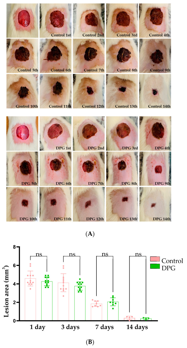
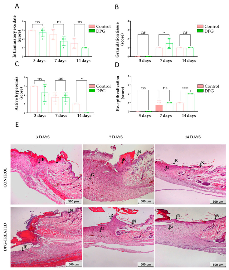
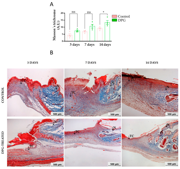
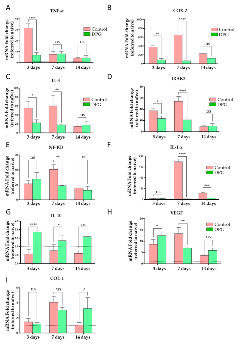
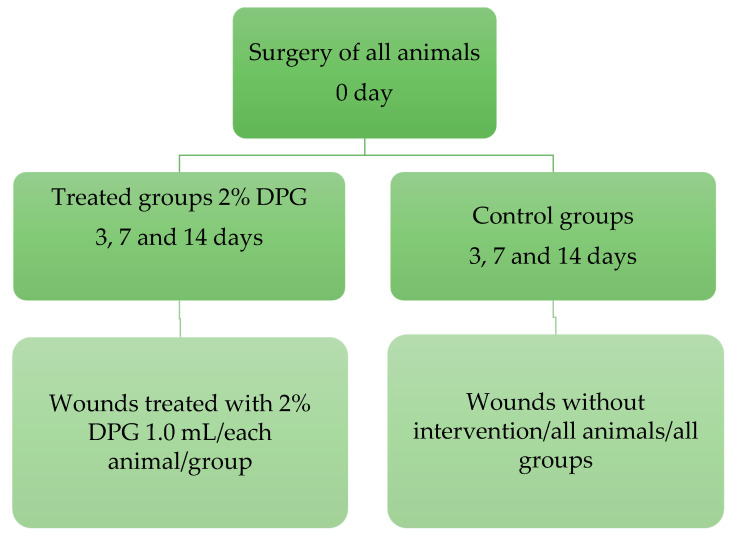
Wound healing is characterized by a systemic and complex process of cellular and molecular activities. Dipotassium Glycyrrhizinate (DPG), a side product derived from glycyrrhizic acid, has several biological effects, such as being antiallergic, antioxidant, antibacterial, antiviral, gastroprotective, antitumoral, and anti-inflammatory. This study aimed to evaluate the anti-inflammatory effect of topical DPG on the healing of cutaneous wounds by secondary intention in an in vivo experimental model. Twenty-four male Wistar rats were used in the experiment, and were randomly divided into six groups of four. Circular excisions were performed and topically treated for 14 days after wound induction. Macroscopic and histopathological analyses were performed. Gene expression was evaluated by real-time qPCR. Our results showed that treatment with DPG caused a decrease in the inflammatory exudate as well as an absence of active hyperemia. Increases in granulation tissue, tissue reepithelization, and total collagen were also observed. Furthermore, DPG treatment reduced the expression of pro-inflammatory cytokines (Tnf-α, Cox-2, Il-8, Irak-2, Nf-kB, and Il-1) while increasing the expression of Il-10, demonstrating anti-inflammatory effects across all three treatment periods. Based on our results, we conclude that DPG attenuates the inflammatory process by promoting skin wound healing through the modulation of distinct mechanisms and signaling pathways, including anti-inflammatory ones. This involves modulation of the expression of pro- and anti-inflammatory cytokine expression; promotion of new granulation tissue; angiogenesis; and tissue re-epithelialization, all of which contribute to tissue remodeling.

Keywords: DPG; animal model; inflammation; rats; skin wound healing.


Conflict of interest statement

The authors declare no conflict of interest.

Figures

Figure 1
Figure 1
(A) Representative macroscopic images of wound healing areas of untreated adult animals (Control—left panel) and those treated with DPG (DPG—right panel) throughout the experimental period (0 to 14 days); (B) Quantitative analysis of the measurements of wound healing areas on days 1, 3, 7, and 14, ns—non significant.
Figure 2
Figure 2
Semi-quantitative characteristics of wound healing areas at days 3, 7, and 14 of experimentation. (A) Inflammatory exudate; (B) active hyperemia, DPG vs. Control, day 14 (* p < 0.05); (C) granulation, DPG vs. Control, day 7 (* p < 0.05); (D) re-epithelialization, DPG vs. Control, day 14 (**** p < 0.0001); (E) representative histological images of the wound healing areas of the animals. Note the presence of normal tissue (N), inflammatory exudate (IE), granulation tissue (G), and re-epithelialization tissue (R). HE staining: 50× magnification (bar = 500 μm).
Figure 3
Figure 3
(A) Quantitative pixel density evaluation of total collagen content in wound scar areas over days 3, 7, and 14 of experimentation, DGP vs. Control, day 14 (* p < 0.05). (B) Representative histological images of the wound scar areas of the animals, both untreated and treated with DPG, throughout the experimentation period. Note that the presence of normal tissue (N) and the presence of total collagen (TC) is highlighted. TM staining: 50× magnification (bar = 500 μm).
Figure 4
Figure 4
Quantitative analysis of the expression of mRNAs in the wound healing samples throughout the days of experimentation in the Control and DPG groups. (A) Tnf-α (**** p < 0.0001); (B) Cox-2 (** p < 0.01, **** p < 0.0001); (C) Il-8 (* p < 0.05, ** p < 0.01); (D) Irak-2 (* p < 0.05, **** p < 0.0001); (E) Nf-kb (** p < 0.01); (F) Il-1α (**** p < 0.0001, *** p < 0.001); (G) Il-10 (**** p < 0.0001, *** p < 0.001); (H) Vegf (* p < 0.05, ** p < 0.01); and (I) Col-1 (* p < 0.05). Ns—not significant, * p < 0.05; ** p < 0.01; *** p < 0.001; **** p < 0.0001 when compared to the control group.
Figure 5
Figure 5
Schematic diagram of the experimental design.

References

    1. Shedoeva A., Leavesley D., Upton Z., Fan C. Wound Healing and the Use of Medicinal Plants. Evid. Based. Complement. Altern. Med. 2019;22:2684108. doi: 10.1155/2019/2684108. - DOI - PMC - PubMed
    1. Rodrigues M., Kosaric N., Bonham C.A., Gurtner G.C. Wound Healing: A Cellular Perspective. Physiol. Rev. 2019;99:665–706. doi: 10.1152/physrev.00067.2017. - DOI - PMC - PubMed
    1. Lindley L.E., Stojadinovic O., Pastar I., Tomic-Canic M. Biology and biomarkers for wound healing. Plast. Reconstr. Surg. 2016;138:18–28. doi: 10.1097/PRS.0000000000002682. - DOI - PMC - PubMed
    1. Scully D., Sfyri P., Wilkinson H.N., Acebes-Huerta A., Verpoorten S., Muñoz-Turrillas M.C., Parnell A., Patel K., Hardman M.J., Gutiérrez L., et al. Optimising platelet secretomes to deliver robust tissue-specific regeneration. J. Tissue Eng. Regen. Med. 2020;14:82–98. doi: 10.1002/term.2965. - DOI - PubMed
    1. Golebiewska E.M., Poole A.W. Platelet secretion: From haemostasis to wound healing and beyond. Blood Rev. 2015;29:153–162. doi: 10.1016/j.blre.2014.10.003. - DOI - PMC - PubMed